


Page 55 - Textbook of Pathology, 6th Edition
P. 55
turns macrophages into foam cells. The examples are as  39
                                                               follows:
                                                               1. Fibrofatty plaques of atherosclerosis (Chapter 15).
                                                               2. Clusters of foam cells in tumour-like masses called
                                                                  xanthomas and xanthelasma.

                                                               Stromal Fatty Infiltration                             CHAPTER 3

                                                               This form of lipid accumulation is quite different from fatty
                                                               change just described. Stromal fatty infiltration is the
                                                               deposition of mature adipose cells in the stromal connective
                                                               tissue in contrast to intracellular deposition of fat in the
                                                               parenchymal cells in fatty change. The condition occurs most
                                                               often in patients with obesity. The two commonly affected
                                                               organs are the heart and the pancreas. Thus, heart can be the
                                                               site for intramyocardial fatty change as well as epicardial
                                                               (stromal) fatty infiltration. The presence of mature adipose
                                                               cells in the stroma generally does not produce any
                                                               dysfunction. In the case of heart, stromal fatty infiltration is
                                                               associated with increased adipose tissue in the epicardium.  Cell Injury and Cellular Adaptations


                                                               INTRACELLULAR ACCUMULATION OF PROTEINS
                                                               Pathologic accumulation of proteins in the cytoplasm of cells
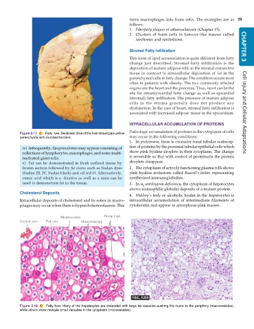
           Figure 3.17  Fatty liver. Sectioned slice of the liver shows pale yellow
           parenchyma with rounded borders.                    may occur in the following conditions:
                                                               1. In proteinuria, there is excessive renal tubular reabsorp-
            iv) Infrequently, lipogranulomas may appear consisting of  tion of proteins by the proximal tubular epithelial cells which
            collections of lymphocytes, macrophages, and some multi-  show pink hyaline droplets in their cytoplasm. The change
            nucleated giant cells.                             is reversible so that with control of proteinuria the protein
            v) Fat can be demonstrated in fresh unfixed tissue by  droplets disappear.
            frozen section followed by fat stains such as Sudan dyes  2. The cytoplasm of actively functioning plasma cells shows
            (Sudan III, IV, Sudan black) and oil red O. Alternatively,  pink hyaline inclusions called Russell’s bodies representing
            osmic acid which is a  fixative as well as a stain can be  synthesised immunoglobulins.
            used to demonstrate fat in the tissue.             3. In α -antitrypsin deficiency, the cytoplasm of hepatocytes
                                                                      1
                                                               shows eosinophilic globular deposits of a mutant protein.
           Cholesterol Deposits
                                                               4. Mallory’s body or alcoholic hyalin in the hepatocytes is
           Intracellular deposits of cholesterol and its esters in macro-  intracellular accumulation of intermediate filaments of
           phages may occur when there is hypercholesterolaemia. This  cytokeratin and appear as amorphous pink masses.


           Figure 3.18  Fatty liver. Many of the hepatocytes are distended with large fat vacuoles pushing the nuclei to the periphery (macrovesicles),
           while others show multiple small vacuoles in the cytoplasm (microvesicles).

