


Page 501 - Textbook of Pathology, 6th Edition
P. 501
485
                                                                 MORPHOLOGIC FEATURES. The disease charac-
                                                                 teristically affects distal bronchi and bronchioles beyond
                                                                 the segmental bronchi.
                                                                 Grossly, the lungs may be involved diffusely or
                                                                 segmentally. Bilateral involvement of lower lobes occurs
                                                                 most frequently. More vertical air passages of left lower
                                                                 lobe are more often involved than the right. The pleura is
                                                                 usually fibrotic and thickened with adhesions to the chest
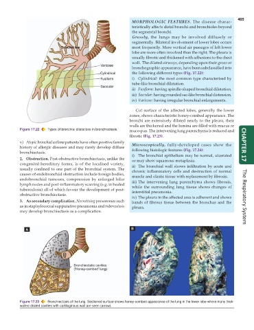
                                                                 wall.  The dilated airways, depending upon their gross or
                                                                 bronchographic appearance, have been subclassified into
                                                                 the following different types (Fig. 17.22):
                                                                 i) Cylindrical: the most common type characterised by
                                                                 tube-like bronchial dilatation.
                                                                 ii) Fusiform: having spindle-shaped bronchial dilatation.
                                                                 iii) Saccular: having rounded sac-like bronchial distension.
                                                                 iv) Varicose: having irregular bronchial enlargements.

                                                                  Cut surface of the affected lobes, generally the lower
                                                               zones, shows characteristic honey-combed appearance. The
                                                               bronchi are extensively dilated nearly to the pleura, their
                                                               walls are thickened and the lumina are filled with mucus or
           Figure 17.22  Types of bronchial dilatations in bronchiectasis.  muco-pus. The intervening lung parenchyma is reduced and
                                                               fibrotic (Fig. 17.23).
           v) Atopic bronchial asthma patients have often positive family
           history of allergic diseases and may rarely develop diffuse  Microscopically, fully-developed cases show the  CHAPTER 17
           bronchiectasis.                                       following histologic features (Fig. 17.24):
                                                                 i) The bronchial epithelium may be normal, ulcerated
           2. Obstruction. Post-obstructive bronchiectasis, unlike the  or may show squamous metaplasia.
           congenital-hereditary forms, is of the localised variety,  ii) The bronchial wall shows infiltration by acute and
           usually confined to one part of the bronchial system. The  chronic inflammatory cells and destruction of normal
           causes of endobronchial obstruction include foreign bodies,  muscle and elastic tissue with replacement by fibrosis.
           endobronchial tumours, compression by enlarged hilar
           lymph nodes and post-inflammatory scarring (e.g. in healed  iii) The intervening lung parenchyma shows fibrosis,
                                                                 while the surrounding lung tissue shows changes of
           tuberculosis) all of which favour the development of post-  interstitial pneumonia.
           obstructive bronchiectasis.
                                                                 iv) The pleura in the affected area is adherent and shows  The Respiratory System
           3. As secondary complication. Necrotising pneumonias such  bands of fibrous tissue between the bronchus and the
           as in staphylococcal suppurative pneumonia and tuberculosis  pleura.
           may develop bronchiectasis as a complication.


           Figure 17.23  Bronchiectasis of the lung. Sectioned surface shows honey-combed appearance of the lung in the lower lobe where many thick-
           walled dilated cavities with cartilaginous wall are seen (arrow).

