


Page 500 - Textbook of Pathology, 6th Edition
P. 500
484 neutrophils. These result in further release of mediators  oedema and inflammatory infiltrate consisting of
           which accentuate the above-mentioned effects. In addition,  lymphocytes and plasma cells with prominence of eosino-
           inflammatory injury is caused by neutrophils and by major  phils. There is hypertrophy of submucosal glands as well
           basic protein (MBP) of eosinophils.                   as of the bronchial smooth muscle.
           2. Intrinsic (idiosyncratic, non-atopic) asthma. This type  4. Changes of bronchitis and emphysema may super-
           of asthma develops later in adult life with negative personal  vene, especially in intrinsic asthma.
           or family history of allergy, negative skin test and normal
           serum levels of IgE. Most of these patients develop typical  CLINICAL FEATURES.  Asthmatic patients suffer from
           symptom-complex after an upper respiratory tract infection  episodes of acute exacerbations interspersed with symptom-
           by viruses. Associated nasal polypi and chronic bronchitis  free periods. Characteristic clinical features are paroxysms
           are commonly present. There are no recognisable allergens  of dyspnoea, cough and wheezing. Most attacks typically
           but about 10% of patients become hypersensitive to drugs,  last for a few minutes to hours. When attacks occur
           most notably to small doses of aspirin (aspirin-sensitive  continuously it may result in more serious condition called
           asthma).                                            status asthmaticus. The clinical diagnosis is supported by
                                                               demonstration of circulation eosinophilia and sputum
           3. Mixed type. Many patients do not clearly fit into either
           of the above two categories and have mixed features of both.  demonstration of Curschmann’s spirals and Charcot-Leyden
           Those patients who develop asthma in early life have strong  crystals. More chronic cases may develop cor pulmonale.
           allergic component, while those who develop the disease late
           tend to be non-allergic. Either type of asthma can be  BRONCHIECTASIS
           precipitated by cold, exercise and emotional stress.  Bronchiectasis is defined as abnormal and irreversible
                                                               dilatation of the bronchi and bronchioles (greater than 2 mm
            MORPHOLOGIC FEATURES. The pathologic changes       in diameter) developing secondary to inflammatory
            are similar in both major types of asthma. The pathologic  weakening of the bronchial walls. The most characteristic
            material examined is generally autopsy of lungs in  clinical manifestation of bronchiectasis is persistent cough
            patients dying of status asthmaticus but the changes are  with expectoration of copious amounts of foul-smelling,
            expected to be similar in non-fatal cases.         purulent sputum. Post-infectious cases commonly develop
            Grossly, the lungs are overdistended due to over-inflation.  in childhood and in early adult life.
            The cut surface shows characteristic occlusion of the  ETIOPATHOGENESIS. The origin of inflammatory destruc-
            bronchi and bronchioles by viscid mucus plugs.     tive process of bronchial walls is nearly always a result of
     SECTION III
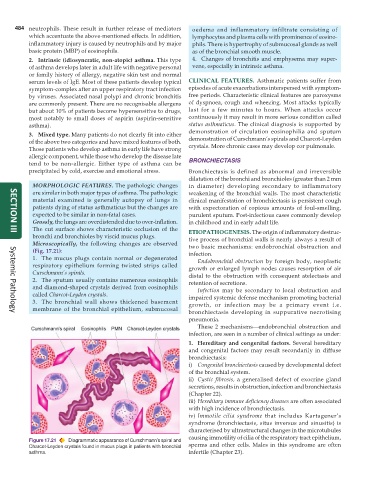
            Microscopically, the following changes are observed  two basic mechanisms: endobronchial obstruction and
            (Fig. 17.21):                                      infection.
            1. The mucus plugs contain normal or degenerated      Endobronchial obstruction by foreign body, neoplastic
            respiratory epithelium forming twisted strips called  growth or enlarged lymph nodes causes resorption of air
            Curschmann’s spirals.                              distal to the obstruction with consequent atelectasis and
            2. The sputum usually contains numerous eosinophils  retention of secretions.
            and diamond-shaped crystals derived from eosinophils  Infection may be secondary to local obstruction and
            called Charcot-Leyden crystals.                    impaired systemic defense mechanism promoting bacterial
            3. The bronchial wall shows thickened basement     growth, or infection may be a primary event i.e.
            membrane of the bronchial epithelium, submucosal
                                                               bronchiectasis developing in suppurative necrotising
     Systemic Pathology
                                                               pneumonia.
                                                                  These 2 mechanisms—endobronchial obstruction and
                                                               infection, are seen in a number of clinical settings as under:
                                                               1. Hereditary and congenital factors. Several hereditary
                                                               and congenital factors may result secondarily in diffuse
                                                               bronchiectasis:
                                                               i) Congenital bronchiectasis caused by developmental defect
                                                               of the bronchial system.
                                                               ii) Cystic fibrosis, a generalised defect of exocrine gland
                                                               secretions, results in obstruction, infection and bronchiectasis
                                                               (Chapter 22).
                                                               iii) Hereditary immune deficiency diseases are often associated
                                                               with high incidence of bronchiectasis.
                                                               iv) Immotile cilia syndrome that includes Kartagener’s
                                                               syndrome (bronchiectasis, situs inversus and sinusitis) is
                                                               characterised by ultrastructural changes in the microtubules
                                                               causing immotility of cilia of the respiratory tract epithelium,
           Figure 17.21  Diagrammatic appearance of Curschmann’s spiral and
           Charcot-Leyden crystals found in mucus plugs in patients with bronchial  sperms and other cells. Males in this syndrome are often
           asthma.                                             infertile (Chapter 23).

