


Page 56 - Textbook of Pathology, 6th Edition
P. 56
40 INTRACELLULAR ACCUMULATION OF GLYCOGEN detected by incubation of tissue section in the solution of
dihydroxy phenyl alanine (DOPA). If the enzyme is present,
Conditions associated with excessive accumulation of dark pigment is identified in pigment cells. This test is called
intracellular glycogen are as under: as DOPA reaction and is particularly useful in differentiating
1. In diabetes mellitus, there is intracellular accumulation of amelanotic melanoma from other anaplastic tumours.
glycogen in different tissues because normal cellular uptake Various disorders of melanin pigmentation cause
of glucose is impaired. Glycogen deposits in diabetes mellitus generalised and localised hyperpigmentation and
are seen in epithelium of distal portion of proximal convolu- hypopigmentation:
ted tubule and descending loop of Henle, in the hepatocytes,
in beta cells of pancreatic islets, and in cardiac muscle cells. i) Generalised hyperpigmentation:
SECTION I
Deposits of glycogen produce clear vacuoles in the cytoplasm a) In Addison’s disease, there is generalised hyper-
of the affected cells. Best’s carmine and periodic acid-Schiff pigmentation of the skin, especially in areas exposed to light,
(PAS) staining may be employed to confirm the presence of and of buccal mucosa.
glycogen in the cells. b) Chloasma observed during pregnancy is the hyper-
2. In glycogen storage diseases or glycogenosis, there is defec- pigmentation on the skin of face, nipples, and genitalia and
tive metabolism of glycogen due to genetic disorders. These occurs under the influence of oestrogen. A similar appear-
conditions along with other similar genetic disorders are ance may be observed in women taking oral contraceptives.
discussed in Chapter 10. c) In chronic arsenical poisoning, there is characteristic rain-
drop pigmentation of the skin.
PIGMENTS ii) Focal hyperpigmentation:
a) Cäfe-au-lait spots are pigmented patches seen in
Pigments are coloured substances present in most living neurofibromatosis and Albright’s syndrome.
beings including humans. There are 2 broad categories of b) Peutz-Jeghers syndrome is characterised by focal peri-oral
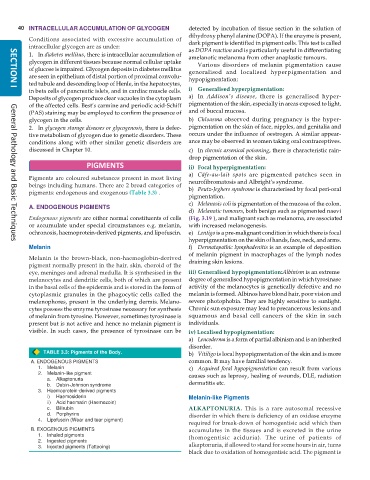
pigments: endogenous and exogenous (Table 3.3) .
pigmentation.
c) Melanosis coli is pigmentation of the mucosa of the colon.
A. ENDOGENOUS PIGMENTS
d) Melanotic tumours, both benign such as pigmented naevi
Endogenous pigments are either normal constituents of cells (Fig. 3.19 ), and malignant such as melanoma, are associated
or accumulate under special circumstances e.g. melanin, with increased melanogenesis.
ochronosis, haemoprotein-derived pigments, and lipofuscin. e) Lentigo is a pre-malignant condition in which there is focal
hyperpigmentation on the skin of hands, face, neck, and arms.
General Pathology and Basic Techniques
Melanin f) Dermatopathic lymphadenitis is an example of deposition
of melanin pigment in macrophages of the lymph nodes
Melanin is the brown-black, non-haemoglobin-derived draining skin lesions.
pigment normally present in the hair, skin, choroid of the
eye, meninges and adrenal medulla. It is synthesised in the iii) Generalised hypopigmentation:Albinism is an extreme
melanocytes and dendritic cells, both of which are present degree of generalised hypopigmentation in which tyrosinase
in the basal cells of the epidermis and is stored in the form of activity of the melanocytes is genetically defective and no
cytoplasmic granules in the phagocytic cells called the melanin is formed. Albinos have blond hair, poor vision and
melanophores, present in the underlying dermis. Melano- severe photophobia. They are highly sensitive to sunlight.
cytes possess the enzyme tyrosinase necessary for synthesis Chronic sun exposure may lead to precancerous lesions and
of melanin from tyrosine. However, sometimes tyrosinase is squamous and basal cell cancers of the skin in such
present but is not active and hence no melanin pigment is individuals.
visible. In such cases, the presence of tyrosinase can be iv) Localised hypopigmentation:
a) Leucoderma is a form of partial albinism and is an inherited
disorder.
TABLE 3.3: Pigments of the Body. b) Vitiligo is local hypopigmentation of the skin and is more
A. ENDOGENOUS PIGMENTS common. It may have familial tendency.
1. Melanin c) Acquired focal hypopigmentation can result from various
2. Melanin-like pigment causes such as leprosy, healing of wounds, DLE, radiation
a. Alkaptonuria
b. Dubin-Johnson syndrome dermatitis etc.
3. Haemoprotein-derived pigments
i) Haemosiderin Melanin-like Pigments
ii) Acid haematin (Haemozoin)
c. Bilirubin ALKAPTONURIA. This is a rare autosomal recessive
d. Porphyrins disorder in which there is deficiency of an oxidase enzyme
4. Lipofuscin (Wear and tear pigment)
required for break-down of homogentisic acid which then
B. EXOGENOUS PIGMENTS accumulates in the tissues and is excreted in the urine
1. Inhaled pigments (homogentisic aciduria). The urine of patients of
2. Ingested pigments
3. Injected pigments (Tattooing) alkaptonuria, if allowed to stand for some hours in air, turns
black due to oxidation of homogentisic acid. The pigment is

