
Page 651 - Textbook of Pathology, 6th Edition
P. 651
635
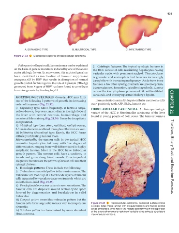
Figure 21.33 Macroscopic patterns of hepatocellular carcinoma.
Pathogenesis of hepatocellular carcinoma can be explained 2 Cytologic features: The typical cytologic features in
on the basis of genetic mutations induced by one of the above the HCC consist of cells resembling hepatocytes having
major etiologic factors. In many cases, this mutated gene has vesicular nuclei with prominent nucleoli. The cytoplasm
been identified as inactivation of tumour suppressor is granular and eosinophilic but becomes increasingly
oncogene p53 by HBV that results in disruption of normal basophilic with increasing malignancy. Aside from these
growth control. In this regards, the role of X-protein (HBxAg) features, a few other cytologic variants are: pleomorphism,
generated from X-gene of HBV has been found to contribute bizarre giant cell formation, spindle-shaped cells, tumour
to carcinogenesis by binding to p53. cells with clear cytoplasm, presence of bile within dilated
canaliculi, and intracytoplasmic Mallory’s hyalin.
MORPHOLOGIC FEATURES. Grossly, HCC may form
one of the following 3 patterns of growth, in decreasing Immunohistochemically, hepatocellular carcinoma cells
order of frequency (Fig. 21.33): stain positively with AFP, EMA, keratin etc. CHAPTER 21
i) Expanding type: Most frequently, it forms a single, FIBROLAMELLAR CARCINOMA. A clinicopathologic
yellow-brown, large mass, most often in the right lobe of variant of the HCC is fibrolamellar carcinoma of the liver
the liver with central necrosis, haemorrhage and found in young people of both sexes. The tumour forms a
occasional bile-staining (Fig. 21.34). It may be deceptively
encapsulated.
ii) Multifocal type: Less often, multifocal, multiple masses,
3-5 cm in diameter, scattered throughout the liver are seen.
iii) Infiltrating (Spreading) type: Rarely, the HCC forms
diffusely infiltrating tumour mass.
Microscopically, the tumour cells in the typical HCC
resemble hepatocytes but vary with the degree of
differentiation, ranging from well-differentiated to highly
anaplastic lesions. Most of the HCC have trabecular
growth pattern. The tumour cells have a tendency to
invade and grow along blood vessels. Thus important The Liver, Biliary Tract and Exocrine Pancreas
diagnostic features are the patterns of tumour cells and their
cytologic features:
1. Histologic patterns: These include the following:
i) Trabecular or sinusoidal pattern is the most common. The
trabeculae are made up of 2-8 cell wide layers of tumour
cells separated by vascular spaces or sinusoids which are
endothelium-lined (Fig. 21.35).
ii) Pseudoglandular or acinar pattern is seen sometimes. The
tumour cells are disposed around central cystic space
formed by degeneration and breakdown in solid
trabeculae.
iii) Compact pattern resembles trabecular pattern but the
tumour cells form large solid masses with inconspicuous Figure 21.34 Hepatocellular carcinoma. Sectioned surface shows
sinusoids. a single, large mass (arrow) with irregular borders and having central
iv) Scirrhous pattern is characterised by more abundant areas of necrosis, while rest of the hepatic parenchyma in the upper part
of the picture shows many nodules of variable sizes owing to co-existent
fibrous stroma. macronodular cirrhosis.