
Page 683 - Textbook of Pathology, 6th Edition
P. 683
is characterised by formation of ‘crescents’ (crescentic GN) 667
outside the glomerular capillaries (extracapillary GN).
‘Crescents’ are formed from the proliferation of parietal
epithelial cells lining Bowman’s capsule with contribution
from visceral epithelial cells and the invading mononuclear
cells. The stimulus for crescent formation appears to be the
presence of fibrin in the capsular space. RPGN occurs most
frequently in adults, with a slight male preponderance.
Prognosis of RPGN in general is dismal.
ETIOPATHOGENESIS. A number of primary glomerular
and systemic diseases are characterised by formation of
crescents. Based on the etiologic agents and pathogenetic
mechanism, patients with RPGN are divided into 3 groups
(Table 22.10):
RPGN in systemic diseases (anti-GBM type);
post-infectious RPGN (immune-complex type); and
pauci-immune RPGN.
Following three serologic markers are used for
categorising RPGN:
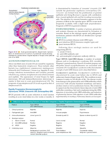
Figure 22.13 Acute glomerulonephritis, diagrammatic represen- i) serum C3 level,
tation of ultrastructure of a portion of glomerular lobule showing ii) anti-GBM antibody; and
characteristic electron-dense irregular deposits or ‘humps’ on the epithelial iii) anti-neutrophil cytoplasmic antibody (ANCA).
side of the GBM.
Type I RPGN: Anti-GBM disease. A number of systemic
ACUTE NON-STREPTOCOCCAL GN diseases such as Goodpasture’s syndrome, SLE, vasculitis,
About one-third cases of acute GN are caused by organisms Wegener’s granulomatosis, Henoch-Schonlein purpura and CHAPTER 22
other than haemolytic streptococci. These include other idiopathic mixed cryoglobulinaemia are associated with
bacteria (e.g. staphylococci, pneumococci, meningococci, crescentic GN. Goodpasture’s syndrome is the characteristic
Salmonella and Pseudomonas), viruses (e.g. hepatitis B virus, example of anti-GBM disease and is described below:
mumps, infectious mononucleosis and varicella), parasitic Goodpasture’s syndrome. Goodpasture’s syndrome is
infections (e.g. malaria, toxoplasmosis and schistosomiasis) characterised by acute renal failure due to RPGN and
and syphilis. The appearance of renal biopsy by light pulmonary haemorrhages (page 494). The condition is more
microscopy, EM and immunofluorescence microscopy is common in males in 3rd decade of life. The disease results
similar to that seen in acute post-streptococcal GN. The from damage to the glomeruli by anti-GBM antibodies which
prognosis of non-streptococcal GN is not as good as that of cross-react with alveolar basement membrane and hence,
streptococcal GN. produce renal as well as pulmonary lesions. The evidences
in support are the characteristic linear deposits of anti-GBM
Rapidly Progressive Glomerulonephritis antibodies consisting of IgG and complement along the GBM,
(Synonyms: RPGN, Crescentic GN, Extracapillary GN) The Kidney and Lower Urinary Tract
detection of circulating anti-GBM antibodies and induction
RPGN presents with an acute reduction in renal function of glomerular lesions with injection of anti-GBM antibodies
resulting in acute renal failure in a few weeks or months. It experimentally in monkeys. Pulmonary lesions can be experi-
TABLE 22.10: Distinguishing Features of Three Main Categories of Rapidly Progressive Glomerulonephritis.
Feature Type I RPGN Type II RPGN Type III RPGN
(Anti-GBM Disease) (Immune Complex Disease) (Pauci-immune GN)
1. Clinical syndrome Nephritic Nephritic Nephritic
2. Pathogenetic type Anti-GBM lmmune-complex Pauci-immune
3. Immunofluorescence Linear Ig and C3 Granular Ig and C3 Sparse or absent Ig and C3
4. Serologic markers
i) Serum C3 level Normal Low-to-normal Normal
ii) Anti-GBM antibody Positive Negative Negative
iii) ANCA Negative Negative Positive
5. Underlying cause Idiopathic Idiopathic Idiopathic
Goodpasture’s syndrome, SLE, Post-infectious Polyarteritis nodosa,
vasculitis, Wegener’s granulomatosis, (post-streptococcal GN) Wegener’s granulomatosis
Henoch-Schonlein purpura