
Page 688 - Textbook of Pathology, 6th Edition
P. 688
672
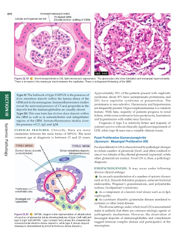
Figure 22.19 Membranoproliferative GN, light microscopic appearance. The glomerular tufts show lobulation and mesangial hypercellularity.
There is increase in the mesangial matrix between the capillaries. There is widespread thickening of the GBM.
Approximately 50% of the patients present with nephrotic
Type II: The hallmark of type II MPGN is the presence of syndrome; about 30% have asymptomatic proteinuria; and
dense amorphous deposits within the lamina densa of the 20% have nephritic syndrome at presentation. The
GBM and in the mesangium. Immunofluorescence studies proteinuria is non-selective. Haematuria and hypertension
reveal the universal presence of C3 and properdin in the
deposits but the immunoglobulins are usually absent. are frequently present. Hypocomplementaemia is a common
Type III: This rare form has electron-dense deposits within feature. With time, majority of patients progress to renal
failure, while some continue to have proteinuria, haematuria
the GBM as well as in subendothelial and subepithelial and hypertension with stable renal function.
SECTION III
regions of the GBM. Immunofluorescence studies show
the presence of C3, IgG and IgM. Prognosis of type I is relatively better and majority of
patients survive without clinically significant impairment of
CLINICAL FEATURES. Clinically, there are many GFR, while type II cases run a variable clinical course.
similarities between the main forms of MPGN. The most
common age at diagnosis is between 15 and 20 years. Focal Proliferative Glomerulonephritis
(Synonym: Mesangial Proliferative GN)
Focal proliferative GN is characterised by pathologic changes
in certain number of glomeruli (focal), and often confined to
one or two lobules of the affected glomeruli (segmental), while
other glomeruli are normal. Focal GN is, thus, a pathologic
Systemic Pathology
diagnosis.
ETIOPATHOGENESIS. It may occur under following
diverse clinical settings:
As an early manifestation of a number of systemic diseases
such as SLE, Henoch-Schonlein purpura, subacute bacterial
endocarditis, Wegener’s granulomatosis, and polyarteritis
nodosa, Goodpasture’s syndrome.
As a component of a known renal disease such as in IgA
nephropathy.
As a primary idiopathic glomerular disease unrelated to
systemic or other renal disease.
The diverse settings under which focal GN is encountered
make it unlikely that there are common etiologic agents or
Figure 22.20 MPGN, diagrammatic representation of ultrastructure pathogenetic mechanisms. However, the observation of
of a portion of glomerular lobule showing features of type I (left half) and mesangial deposits of immunoglobulins and complement
type II (right half) MPGN. Type I (classic form) shows the characteristic suggest immune complex disease and participation of the
subendothelial electron-dense deposit s, while type II (dense deposit
disease) is characterised by intramembranous dense deposit s. mesangium.