
Page 684 - Textbook of Pathology, 6th Edition
P. 684
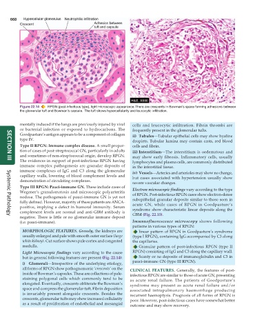
668


           Figure 22.14  RPGN (post-infectious type), light microscopic appearance. There are crescents in Bowman’s space forming adhesions between
           the glomerular tuft and Bowman’s capsule. The tuft shows hypercellularity and leucocytic infiltration.


           mentally induced if the lungs are previously injured by viral  cells and leucocytic infiltration. Fibrin thrombi are
           or bacterial infection or exposed to hydrocarbons. The  frequently present in the glomerular tufts.
           Goodpasture’s antigen appears to be a component of collagen  ii) Tubules—Tubular epithelial cells may show hyaline
           type IV.
                                                                 droplets. Tubular lumina may contain casts, red blood
           Type II RPGN: Immune complex disease. A small propor-  cells and fibrin.
           tion of cases of post-streptococcal GN, particularly in adults  iii) Interstitium—The interstitium is oedematous and
           and sometimes of non-streptococcal origin, develop RPGN.  may show early fibrosis. Inflammatory cells, usually
           The evidences in support of post-infectious RPGN having  lymphocytes and plasma cells, are commonly distributed
     SECTION III
           immune complex pathogenesis are granular deposits of  in the interstitial tissue.
           immune complexes of IgG and C3 along the glomerular   iv) Vessels—Arteries and arterioles may show no change,
           capillary walls, lowering of blood complement levels and  but cases associated with hypertension usually show
           demonstration of circulating complexes.               severe vascular changes.
           Type III RPGN: Pauci-immune GN. These include cases of  Electron microscopic findings vary according to the type
           Wegener’s granulomatosis and microscopic polyarteritis  of RPGN. Post-infectious RPGN cases show electron-dense
           nodosa. The pathogenesis of pauci-immune GN is yet not  subepithelial granular deposits similar to those seen in
           fully defined. However, majority of these patients are ANCA-  acute GN, while cases of RPGN in Goodpasture’s
           positive, implying a defect in humoral immunity. Serum  syndrome show characteristic linear deposits along the
           complement levels are normal and anti-GBM antibody is  GBM (Fig. 22.15).
           negative. There is little or no glomerular immune deposit
     Systemic Pathology
           (i.e. pauci-immune).                                  Immunofluorescence microscopy shows following
                                                                 patterns in various types of RPGN:
            MORPHOLOGIC FEATURES. Grossly, the kidneys are          linear pattern of RPGN in Goodpasture’s syndrome
            usually enlarged and pale with smooth outer surface (large  (type I RPGN), containing IgG accompanied by C3 along
            white kidney). Cut surface shows pale cortex and congested  the capillaries.
            medulla.                                                Granular pattern of post-infectious RPGN (type II
            Light Microscopic findings vary according to the cause  RPGN) consisting of IgG and C3 along the capillary wall.
            but in general following features are present (Fig. 22.14):  Scanty or no deposits of immunoglobulin and C3 in
                                                                 pauci-immune GN (type III RPGN).
            i) Glomeruli—Irrespective of the underlying etiology,
            all forms of RPGN show pathognomonic ‘crescents’ on the  CLINICAL FEATURES.  Generally, the features of post-
            inside of Bowman’s capsules. These are collections of pale-  infectious RPGN are similar to those of acute GN, presenting
            staining polygonal cells which commonly tend to be  as acute renal failure. The patients of Goodpasture’s
            elongated. Eventually, crescents obliterate the Bowman’s  syndrome may present as acute renal failure and/or
            space and compress the glomerular tuft. Fibrin deposition  associated intrapulmonary haemorrhage producing
            is invariably present alongside crescents. Besides the  recurrent haemoptysis. Prognosis of all forms of RPGN is
            crescents, glomerular tufts may show increased cellularity  poor. However, post-infectious cases have somewhat better
            as a result of proliferation of endothelial and mesangial  outcome and may show recovery.