
Page 686 - Textbook of Pathology, 6th Edition
P. 686
670
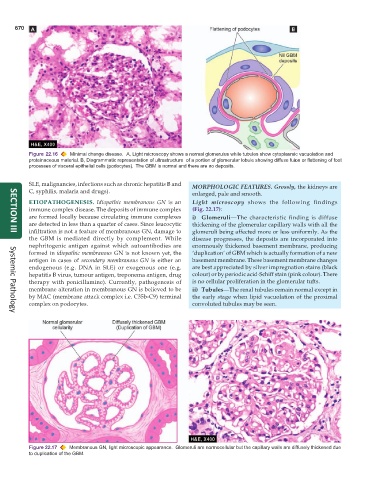
Figure 22.16 Minimal change disease. A, Light microscopy shows a normal glomerulus while tubules show cytoplasmic vacuolation and
proteinaceous material. B, Diagrammatic representation of ultrastructure of a portion of glomerular lobule showing diffuse fusion or flattening of foot
processes of visceral epithelial cells (podocytes). The GBM is normal and there are no deposits.
SLE, malignancies, infections such as chronic hepatitis B and MORPHOLOGIC FEATURES. Grossly, the kidneys are
C, syphilis, malaria and drugs). enlarged, pale and smooth.
ETIOPATHOGENESIS. Idiopathic membranous GN is an Light microscopy shows the following findings
immune complex disease. The deposits of immune complex (Fig. 22.17):
are formed locally because circulating immune complexes i) Glomeruli—The characteristic finding is diffuse
are detected in less than a quarter of cases. Since leucocytic thickening of the glomerular capillary walls with all the
infiltration is not a feature of membranous GN, damage to glomeruli being affected more or less uniformly. As the
SECTION III
the GBM is mediated directly by complement. While disease progresses, the deposits are incorporated into
nephritogenic antigen against which autoantibodies are enormously thickened basement membrane, producing
formed in idiopathic membranous GN is not known yet, the ‘duplication’ of GBM which is actually formation of a new
antigen in cases of secondary membranous GN is either an basement membrane. These basement membrane changes
endogenous (e.g. DNA in SLE) or exogenous one (e.g. are best appreciated by silver impregnation stains (black
hepatitis B virus, tumour antigen, treponema antigen, drug colour) or by periodic acid-Schiff stain (pink colour). There
therapy with penicillamine). Currently, pathogenesis of is no cellular proliferation in the glomerular tufts.
membrane alteration in membranous GN is believed to be ii) Tubules—The renal tubules remain normal except in
by MAC (membrane attack complex i.e. C35b-C9) terminal the early stage when lipid vacuolation of the proximal
complex on podocytes. convoluted tubules may be seen.
Systemic Pathology
Figure 22.17 Membranous GN, light microscopic appearance. Glomeruli are normocellular but the capillary walls are diffusely thickened due
to duplication of the GBM.