
Page 687 - Textbook of Pathology, 6th Edition
P. 687
increase in cellularity of the mesangium associated with 671
increased lobulation of the tuft, and irregular thickening of
the capillary wall.
ETIOPATHOGENESIS. Etiology of MPGN is unknown
though in some cases there is evidence of preceding
streptococcal infection. Based on ultrastructural,
immunofluorescence and pathogenetic mechanisms, three
types of MPGN are recognised:
Type I or classic form is an example of immune complex
disease and comprises more than 70% cases. It is charac-
terised by immune deposits in the subendothelial position.
Immune-complex MPGN is seen in association with systemic
immune-complex diseases (e.g. SLE, mixed cryoglo-
bulinaemia, Sjögren’s syndrome), chronic infections (e.g.
bacterial endocarditis, HIV, hepatitis B and C) and
malignancies (e.g. lymphomas and leukaemias).
Type II or dense deposit disease is the example of alter-
nate pathway disease (page 664) and constitutes about 30%
cases. The capillary wall thickening is due to the deposition
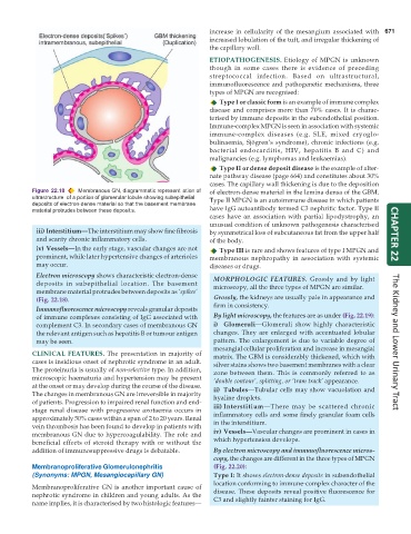
Figure 22.18 Membranous GN, diagrammatic represent ation of of electron-dense material in the lamina densa of the GBM.
ultrastructure of a portion of glomerular lobule showing subepithelial Type II MPGN is an autoimmune disease in which patients
deposits of electron-dense material so that the basement membrane
material protrudes between these deposit s. have IgG autoantibody termed C3 nephritic factor. Type II
cases have an association with partial lipodystrophy, an
unusual condition of unknown pathogenesis characterised
iii) Interstitium—The interstitium may show fine fibrosis by symmetrical loss of subcutaneous fat from the upper half
and scanty chronic inflammatory cells. of the body. CHAPTER 22
iv) Vessels—In the early stage, vascular changes are not Type III is rare and shows features of type I MPGN and
prominent, while later hypertensive changes of arterioles membranous nephropathy in association with systemic
may occur. diseases or drugs.
Electron microscopy shows characteristic electron-dense MORPHOLOGIC FEATURES. Grossly and by light
deposits in subepithelial location. The basement microscopy, all the three types of MPGN are similar.
membrane material protrudes between deposits as ‘spikes’
(Fig. 22.18). Grossly, the kidneys are usually pale in appearance and
Immunofluorescence microscopy reveals granular deposits firm in consistency.
of immune complexes consisting of IgG associated with By light microscopy, the features are as under (Fig. 22.19):
complement C3. In secondary cases of membranous GN i) Glomeruli—Glomeruli show highly characteristic
the relevant antigen such as hepatitis B or tumour antigen changes. They are enlarged with accentuated lobular
may be seen. pattern. The enlargement is due to variable degree of The Kidney and Lower Urinary Tract
mesangial cellular proliferation and increase in mesangial
CLINICAL FEATURES. The presentation in majority of matrix. The GBM is considerably thickened, which with
cases is insidious onset of nephrotic syndrome in an adult. silver stains shows two basement membranes with a clear
The proteinuria is usually of non-selective type. In addition, zone between them. This is commonly referred to as
microscopic haematuria and hypertension may be present ‘double contour’, splitting, or ‘tram track’ appearance.
at the onset or may develop during the course of the disease. ii) Tubules—Tubular cells may show vacuolation and
The changes in membranous GN are irreversible in majority hyaline droplets.
of patients. Progression to impaired renal function and end-
stage renal disease with progressive azotaemia occurs in iii) Interstitium—There may be scattered chronic
approximately 50% cases within a span of 2 to 20 years. Renal inflammatory cells and some finely granular foam cells
vein thrombosis has been found to develop in patients with in the interstitium.
membranous GN due to hypercoagulability. The role and iv) Vessels—Vascular changes are prominent in cases in
beneficial effects of steroid therapy with or without the which hypertension develops.
addition of immunosuppressive drugs is debatable. By electron microscopy and immunofluorescence micros-
copy, the changes are different in the three types of MPGN
Membranoproliferative Glomerulonephritis (Fig. 22.20):
(Synonyms: MPGN, Mesangiocapillary GN) Type I: It shows electron-dense deposits in subendothelial
location conforming to immune-complex character of the
Membranoproliferative GN is another important cause of
nephrotic syndrome in children and young adults. As the disease. These deposits reveal positive fluorescence for
name implies, it is characterised by two histologic features— C3 and slightly fainter staining for IgG.