
Page 711 - Textbook of Pathology, 6th Edition
P. 711
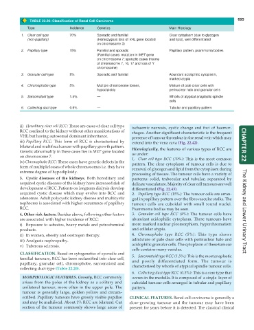
TABLE 22.20: Classification of Renal Cell Carcinoma 695
Type Incidence Genetics Main Histology
1. Clear cell type 70% Sporadic and familial Clear cytoplasm (due to glycogen
(non-papillary) (Homozygous loss of VHL gene located and lipid), well differentiated
on chromosome 3)
2. Papillary type 15% Familial and sporadic Papillary pattern, psammoma bodies
(Familial cases: mutation in MET gene
on chromosome 7; sporadic cases trisomy
of chromosome 7, 16, 17 and loss of Y
chromosome)
3. Granular cell type 8% Sporadic and familial Abundant acidophilic cytoplasm,
marked atypia
4. Chromophobe type 5% Multiple chromosome losses, Mixture of pale clear cells with
hypodiploidy perinuclear halo and granular cells
5. Sarcomatoid type 1.5% — Whorls of atypical anaplastic spindle
cells
6. Collecting duct type 0.5% — Tubular and papillary pattern
ii) Hereditary clear cell RCC: These are cases of clear cell type ischaemic necrosis, cystic change and foci of haemor-
RCC confined to the kidney without other manifestations of rhages. Another significant characteristic is the frequent
VHL but having autosomal dominant inheritance. presence of tumour thrombus in the renal vein which may
iii) Papillary RCC: This form of RCC is characterised by extend into the vena cava (Fig. 22.42).
bilateral and multifocal cancer with papillary growth pattern. CHAPTER 22
Genetic abnormality in these cases lies in MET gene located Histologically, the features of various types of RCC are
on chromosome 7. as under:
iv) Chromophobe RCC: These cases have genetic defects in the 1. Clear cell type RCC (70%): This is the most common
pattern. The clear cytoplasm of tumour cells is due to
form of multiple losses of whole chromosomes i.e. they have removal of glycogen and lipid from the cytoplasm during
extreme degree of hypodiploidy.
processing of tissues. The tumour cells have a variety of
3. Cystic diseases of the kidneys. Both hereditary and patterns: solid, trabecular and tubular, separated by
acquired cystic diseases of the kidney have increased risk of delicate vasculature. Majority of clear cell tumours are well
development of RCC. Patients on longterm dialysis develop differentiated (Fig. 22.43).
acquired cystic disease which may evolve into RCC and 2. Papillary type RCC (15%): The tumour cells are arran-
adenomas. Adult polycystic kidney disease and multicystic ged in papillary pattern over the fibrovascular stalks. The
nephroma is associated with higher occurrence of papillary tumour cells are cuboidal with small round nuclei.
RCC. Psammoma bodies may be seen.
4. Other risk factors. Besides above, following other factors 3. Granular cell type RCC (8%): The tumour cells have The Kidney and Lower Urinary Tract
are associated with higher incidence of RCC: abundant acidophilic cytoplasm. These tumours have
i) Exposure to asbestos, heavy metals and petrochemical more marked nuclear pleomorphism, hyperchromatism
products. and cellular atypia.
ii) In women, obesity and oestrogen therapy. 4. Chromophobe type RCC (5%): This type shows
iii) Analgesic nephropathy. admixture of pale clear cells with perinuclear halo and
v) Tuberous sclerosis. acidophilic granular cells. The cytoplasm of these tumour
cells contains many vesicles.
CLASSIFICATION. Based on cytogenetics of sporadic and
familial tumours, RCC has been reclassified into clear cell, 5. Sarcomatoid type RCC (1.5%): This is the most anaplastic
papillary, granular cell, chromophobe, sarcomatoid and and poorly differentiated form. The tumour is
collecting duct type (Table 22.20). characterised by whorls of atypical spindle tumour cells.
6. Collecting duct type RCC (0.5%): This is a rare type that
MORPHOLOGIC FEATURES. Grossly, RCC commonly occurs in the medulla. It is composed of a single layer of
arises from the poles of the kidney as a solitary and cuboidal tumour cells arranged in tubular and papillary
unilateral tumour, more often in the upper pole. The pattern.
tumour is generally large, golden yellow and circum-
scribed. Papillary tumours have grossly visible papillae CLINICAL FEATURES. Renal cell carcinoma is generally a
and may be multifocal. About 1% RCC are bilateral. Cut slow-growing tumour and the tumour may have been
section of the tumour commonly shows large areas of present for years before it is detected. The classical clinical