
Page 709 - Textbook of Pathology, 6th Edition
P. 709
693
TABLE 22.18: Urinary Calculi (continued)
Morphology
Calcium oxalate stones
Struvite (‘Staghorn’) stone
Uric acid stones
Cystine stones
Figure facing Table 22.18
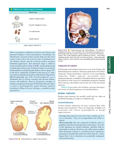
Figure 22.41 Hydronephrosis with nephrolithiasis. The kidney is
there is progressive dilatation of pelvis and calyces and enlarged and heavy. On cut section, the renal pelvis and calyces are
pressure atrophy of renal parenchyma. Eventually, the dilated and cystic and contain a large stone in the pelvis of the kidney
(arrow). The cystic change is seen to extend into renal p
arenchyma,
dilated pelvi-calyceal system extends deep into the renal compressing the cortex as a thin rim at the periphery . Unlike polycystic
cortex so that a thin rim of renal cortex is stretched over kidney, however, these cysts are communicating with the pelvi-calyceal
the dilated calyces and the external surface assumes system.
lobulated appearance. This advanced stage is called as
intrarenal hydronephrosis (Fig. 22.40,B). An important point TUMOURS OF KIDNEY CHAPTER 22
of distinction between the sectioned surface of advanced
hydronephrosis and polycystic kidney disease (page 657) Both benign and malignant tumours occur in the kidney, the
is the direct continuity of dilated cystic spaces (i.e. dila- latter being more common. These may arise from renal tubules
ted calyces) with the renal pelvis in the former (Fig. 22.41). (adenoma, adenocarcinoma), embryonic tissue (mesoblastic
Microscopically, the wall of hydronephrotic sac is nephroma, Wilms’ tumour), mesenchymal tissue
thickened due to fibrous scarring and chronic inflam- (angiomyolipoma, medullary interstitial tumour) and from
matory cell infiltrate. There is progressive atrophy of the epithelium of the renal pelvis (urothelial carcinoma). Besides
tubules and glomeruli alongwith interstitial fibrosis. Stasis these tumours, the kidney may be the site of the secondary
tumours.
of urine in hydronephrosis causes infection (pyelitis) Table 22.19 provides a list of kidney tumours; the impor-
resulting in filling of the sac with pus, a condition called tant forms of renal neoplasms are described below.
pyonephrosis.
BENIGN TUMOURS
Benign renal tumours are usually small and are often an The Kidney and Lower Urinary Tract
incidental finding at autopsy or nephrectomy.
Cortical Adenoma
Cortical tubular adenomas are more common than other
benign renal neoplasms. They are frequently multiple and
associated with chronic pyelonephritis or benign
nephrosclerosis.
Grossly, these tumours may form tiny nodules up to 3
cm in diameter. They are encapsulated and white or
yellow.
Microscopically, they are composed of tubular cords or
papillary structures projecting into cystic space. The cells
of the adenoma are usually uniform, cuboidal with no
atypicality or mitosis. However, size of the tumour rather
than histologic criteria is considered more significant
parameter to predict the behaviour of the tumour—those
larger than 3 cm in diameter are potentially malignant
Figure 22.40 Hydronephrosis, stages in its evolution. and metastasising.