
Page 707 - Textbook of Pathology, 6th Edition
P. 707
Pathogenesis. The mechanism of calcium stone formation is 691
explained on the basis of imbalance between the degree of
supersaturation of the ions forming the stone and the
concentration of inhibitors in the urine. Most likely site where
the crystals of calcium oxalate and/or calcium phosphate
are precipitated is the tubular lining or around some
fragment of debris in the tubule acting as nidus of the stone.
The stone grows, as more and more crystals are deposited
around the nidus. A number of other predisposing factors
contributing to formation of calcium stones are alkaline
urinary pH, decreased urinary volume and increased
excretion of oxalate and uric acid.
Morphology. Calcium stones are usually small (less than a
centimeter), ovoid, hard, with granular rough surface. They
are dark brown due to old blood pigment deposited in them
as a result of repeated trauma caused to the urinary tract by
these sharp-edged stones.
2. MIXED (STRUVITE) STONES. About 15% of urinary
calculi are made of magnesium-ammonium-calcium
phosphate, often called struvite; hence mixed stones are also
called as ‘struvite stones’ or ‘triple phosphate stones’.
Etiology. Struvite stones are formed as a result of infection
of the urinary tract with urea-splitting organisms that
produce urease such as by species of Proteus, and occasionally
Klebsiella, Pseudomonas and Enterobacter. These are, therefore, CHAPTER 22
also known as infection-induced stones. However, E. coli does
not form urease.
Figure 22.38 Causes of obstructive uropathy. Morphology. Struvite stones are yellow-white or grey. They
tend to be soft and friable and irregular in shape. ‘Staghorn
stone’ which is a large, solitary stone that takes the shape of
(renal colic) as they pass down along the ureter and manifest the renal pelvis where it is often formed is an example of
by haematuria. struvite stone (Fig. 22.39).
Types of Urinary Calculi
There are 4 main types of urinary calculi—calcium
containing, mixed (struvite), uric acid and cystine stones, and
a few rare types (Table 22.18). The Kidney and Lower Urinary Tract
1. CALCIUM STONES. Calcium stones are the most
common comprising about 75% of all urinary calculi. They
may be pure stones of calcium oxalate (50%) or calcium
phosphate (5%), or mixture of calcium oxalate and calcium
phosphate (45%).
Etiology. Etiology of calcium stones is variable.
i) About 50% of patients with calcium stones have idiopathic
hypercalciuria without hypercalcaemia.
ii) Approximately 10% cases are associated with hyper-
calcaemia and hypercalciuria, most commonly due to hyper-
parathyroidism, or a defect in the bowel (i.e. absorptive
hypercalciuria), or in the kidney (i.e. renal hypercalciuria).
iii) About 15% of patients with calcium stones have
hyperuricosuria with a normal blood uric acid level and without
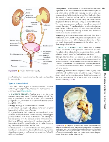
any abnormality of calcium metabolism. Figure 22.39 Staghorn renal stone with chronic pyelonephritis.The
iv) In about 25% of patients with calcium stones, the cause kidney is enlarged and heavy. The capsule is adherent to the cortex and
is unknown as there is no abnormality in urinary excretion has irregular scars on the surface. Sectioned surface shows dilated pelvi-
calyceal system with atrophied and thin peripheral cortex. The pelvis of
of calcium, uric acid or oxalate and is referred to as ‘idiopathic the kidney contains a single, large, sof t yellow white stone t aking the
calcium stone disease’. contour of the pelvi-calyceal system (arrow).