
Page 708 - Textbook of Pathology, 6th Edition
P. 708
692
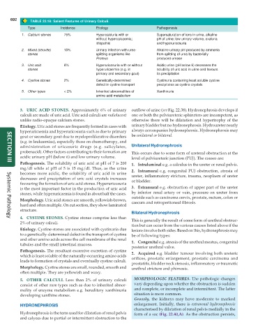
TABLE 22.18: Salient Features of Urinary Calculi.
Type Incidence Etiology Pathogenesis
1. Calcium stones 75% Hypercalciuria with or Supersaturation of ions in urine, alkaline
without hypercalcaemia; pH of urine; low urinary volume, oxaluria
idiopathic and hyperuricosuria
2. Mixed (struvite) 15% Urinary infection with urea- Alkaline urinary pH produced by ammonia
stones splitting organisms like from splitting of urea by bacterially
Proteus produced urease
3. Uric acid 6% Hyperuricosuria with or without Acidic urine (pH below 6) decreases the
stones hyperuricaemia (e.g. in solubility of uric acid in urine and favours
primary and secondary gout) its precipitation
4. Cystine stones 2% Genetically-determined Cystinuria containing least soluble cystine
defect in cystine transport precipitates as cystine crystals
5. Other types < 2% Inherited abnormalities of Xanthinuria
amino acid metabolism
3. URIC ACID STONES. Approximately 6% of urinary outflow of urine (see Fig. 22.38). Hydronephrosis develops if
calculi are made of uric acid. Uric acid calculi are radiolucent one or both the pelviureteric sphincters are incompetent, as
unlike radio-opaque calcium stones. otherwise there will be dilatation and hypertrophy of the
Etiology. Uric acid stones are frequently formed in cases with urinary bladder but no hydronephrosis. Hydroureter nearly
hyperuricaemia and hyperuricosuria such as due to primary always accompanies hydronephrosis. Hydronephrosis may
gout or secondary gout due to myeloproliferative disorders be unilateral or bilateral.
(e.g. in leukaemias), especially those on chemotherapy, and
administration of uricosuric drugs (e.g. salicylates, Unilateral Hydronephrosis
probenacid). Other factors contributing to their formation are This occurs due to some form of ureteral obstruction at the
acidic urinary pH (below 6) and low urinary volume. level of pelviureteric junction (PUJ). The causes are:
Pathogenesis. The solubility of uric acid at pH of 7 is 200 1. Intraluminal e.g. a calculus in the ureter or renal pelvis.
SECTION III
mg/dl while at pH of 5 is 15 mg/dl. Thus, as the urine 2. Intramural e.g. congenital PUJ obstruction, atresia of
becomes more acidic, the solubility of uric acid in urine ureter, inflammatory stricture, trauma, neoplasm of ureter
decreases and precipitation of uric acid crystals increases or bladder.
favouring the formation of uric acid stones. Hyperuricosuria
is the most important factor in the production of uric acid 3. Extramural e.g. obstruction of upper part of the ureter
stones, while hyperuricaemia is found in about half the cases. by inferior renal artery or vein, pressure on ureter from
outside such as carcinoma cervix, prostate, rectum, colon or
Morphology. Uric acid stones are smooth, yellowish-brown, caecum and retroperitoneal fibrosis.
hard and often multiple. On cut section, they show laminated
structure.
Bilateral Hydronephrosis
4. CYSTINE STONES. Cystine stones comprise less than This is generally the result of some form of urethral obstruc-
Systemic Pathology
2% of urinary calculi.
tion but can occur from the various causes listed above if the
Etiology. Cystine stones are associated with cystinuria due lesions involve both sides. Based on this, hydronephrosis may
to a genetically-determined defect in the transport of cystine be of following types:
and other amino acids across the cell membrane of the renal 1. Congenital e.g. atresia of the urethral meatus, congenital
tubules and the small intestinal mucosa.
posterior urethral valve.
Pathogenesis. The resultant excessive excretion of cystine 2. Acquired e.g. bladder tumour involving both ureteric
which is least soluble of the naturally-occurring amino acids orifices, prostatic enlargement, prostatic carcinoma and
leads to formation of crystals and eventually cystine calculi.
prostatitis, bladder neck stenosis, inflammatory or traumatic
Morphology. Cystine stones are small, rounded, smooth and urethral stricture and phimosis.
often multiple. They are yellowish and waxy.
5. OTHER CALCULI. Less than 2% of urinary calculi MORPHOLOGIC FEATURES. The pathologic changes
consist of other rare types such as due to inherited abnor- vary depending upon whether the obstruction is sudden
mality of enzyme metabolism e.g. hereditary xanthinuria and complete, or incomplete and intermittent. The latter
developing xanthine stones. situation is more common.
Grossly, the kidneys may have moderate to marked
HYDRONEPHROSIS enlargement. Initially, there is extrarenal hydronephrosis
characterised by dilatation of renal pelvis medially in the
Hydronephrosis is the term used for dilatation of renal pelvis form of a sac (Fig. 22.40,A). As the obstruction persists,
and calyces due to partial or intermittent obstruction to the