
Page 706 - Textbook of Pathology, 6th Edition
P. 706
690
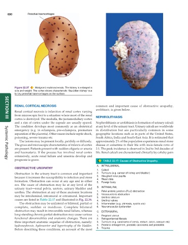
Figure 22.37 Malignant nephrosclerosis. The kidney is enlarged in
size and weight. The cortex shows characteristic ‘flea bitten kidney’ due
to tiny petechial haemorrhages on the surface.
RENAL CORTICAL NECROSIS common and important cause of obstructive uropathy,
urolithiasis, is given below.
Renal cortical necrosis is infarction of renal cortex varying
from microscopic foci to a situation where most of the renal NEPHROLITHIASIS
cortex is destroyed. The medulla, the juxtamedullary cortex
SECTION III
and a rim of cortex under the capsule are usually spared. Nephrolithiasis or urolithiasis is formation of urinary calculi
The condition develops most commonly as an obstetrical at any level of the urinary tract. Urinary calculi are worldwide
emergency (e.g. in eclampsia, pre-eclampsia, premature in distribution but are particularly common in some
separation of the placenta). Other causes include septic shock, geographic locations such as in parts of the United States,
poisoning, severe trauma etc. South Africa, India and South-East Asia. It is estimated that
The lesions may be present focally, patchily or diffusely. approximately 2% of the population experiences renal stone
The gross and microscopic characteristics of infarcts of cortex disease at sometime in their life with male-female ratio of
are present. Patients present with sudden oliguria or anuria 2:1. The peak incidence is observed in 2nd to 3rd decades of
and haematuria. If the process has involved renal cortex life. Renal calculi are characterised clinically by colicky pain
extensively, acute renal failure and uraemia develop and
prognosis is grave. TABLE 22.17: Causes of Obstructive Uropathy.
Systemic Pathology
A. INTRALUMINAL
OBSTRUCTIVE UROPATHY
1. Calculi
Obstruction in the urinary tract is common and important 2. Tumours (e.g. cancer of kidney and bladder)
because it increases the susceptibility to infection and stone 3. Sloughed renal papilla
formation. Obstruction can occur at any age and in either 4. Blood clots
Foreign body
5.
sex. The cause of obstruction may lie at any level of the
urinary tract—renal pelvis, ureters, urinary bladder and B. INTRAMURAL
urethra. The obstruction at any of these anatomic locations 1. Pelvi-ureteric junction (PUJ) obstruction
may be intraluminal, intramural or extramural. Important 2. Vesicoureteric obstruction
Urethral stricture
3.
causes are listed in Table 22.17 and illustrated in Fig. 22.38. 4. Urethral valves
The obstruction may be unilateral or bilateral, partial or 5. Inflammation (e.g. phimosis, cystitis etc)
complete, sudden or insidious. Complete bilateral 6. Neuromuscular dysfunction
obstruction may result in irreversible renal failure, whereas C. EXTRAMURAL
long-standing chronic partial obstruction may cause various 1. Pregnant uterus
functional abnormalities and anatomic changes. There are 2. Retroperitoneal fibrosis
three important anatomic sequelae of obstruction, namely: 3. Tumours (e.g. carcinoma of cervix, rectum, colon, caecum etc)
hydronephrosis, hydroureter and hypertrophy of the bladder. 4. Prostatic enlargement, prostatic carcinoma and prostatitis
Before describing these conditions, an account of the most 5. Trauma