
Page 743 - Textbook of Pathology, 6th Edition
P. 743
727
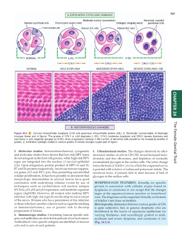
Figure 24.5 Cervical intraepithelial neoplasia (CIN) and squamous intraepithelial lesions (SIL). A, Schematic representation of histologic CHAPTER 24
changes (lower part of figure). The grades of CIN-1 or mild dysplasia (L-SIL), CIN-2 (moderate dysplasia) and CIN-3 (severe dysplasia and
carcinoma in situ) (together grouped as H-SIL) show progressive increase in the number of abnormal cells parallel to the increasing severity of
grades. B, Exfoliative cytologic studies in various grades of cellular changes (upper part of figure). The Female Genital Tract
3. Molecular studies. Immunohistochemical, cytogenetic 5. Ultrastructural studies. The changes observed on ultra-
and molecular studies have shown that low-risk HPV types structural studies of cells in CIN/SIL reveal increased mito-
do not integrate in the host cell genome, while high-risk HPV chondria and free ribosomes, and depletion of normally
types are integrated into the nucleus of cervical epithelial accumulated glycogen in the surface cells. The latter change
cells. Upon integration, protein product of HPV-16 and 18, forms the basis of Schiller’s test in which the suspected cervix
E7 and E6 proteins respectively, inactivate tumour suppres- is painted with solution of iodine and potassium iodide. The
sor genes, p53 and RB-1 gene, thus permitting uncontrolled cancerous focus, if present, fails to stain because of lack of
cellular proliferation. It has been possible to document that glycogen in the surface cells.
morphologic abnormalities in cervical lesions have good
correlation with underlying cellular events by use of MORPHOLOGIC FEATURES. Grossly, no specific
techniques such as proliferation cell nuclear antigen picture is associated with cellular atypia found in
(PCNA), p16, p53 and p63 expression, and nucleolar organizer dysplasias or carcinoma in situ except that the changes
region (AgNOR). However, all women who harbour HPV begin at the squamocolumnar junction or transitional
infection with high-risk type do not develop invasive cancer zone. The diagnosis can be suspected clinically on the basis
of the cervix. Women who have persistence of this infection of Schiller’s test done on bedside.
or those who have another cofactor such as cigarette smoking Histologically, distinction between various grades of CIN
or immunodeficiency, are at greater risk to develop is quite subjective, but, in general dysplastic cells are
progression of lesions. distributed in the layers of squamous epithelium for
4. Immunologic studies. Circulating tumour specific anti- varying thickness, and accordingly graded as mild,
gens and antibodies are detected in patients of cervical cancer. moderate and severe dysplasia, and carcinoma in situ
Antibodies to virus specific antigens are identified on tumour (Fig. 24.5,A).
cells and in sera of such patients.