
Page 744 - Textbook of Pathology, 6th Edition
P. 744
728 In mild dysplasia (CIN-1), the abnormal cells extend up worldwide cervical cancer remains third most common
to one-third thickness from the basal to the surface layer; cancer in women, next to breast and lung cancer. Although
In moderate dysplasia (CIN-2) up to two-thirds; accurate statistics are not available from India, but it is
In severe dysplasia (CIN-3), these cells extend from 75- perhaps the leading cause of death in women. In the Pap
90% thickness of epithelium; and screening programme, patients having abnormal Pap smear
In carcinoma in situ (included in CIN-3), the entire are appropriately followed up and, therefore, it requires
thickness from the basement membrane to the surface understanding of the Bethesda system by the clinician as
shows dysplastic cells. regards value and limitations of cytology reports prepared
The atypical cells migrate to the surface layers from by the cytologist/cytotechnician.
where they are shed off (exfoliated) into vaginal secre- Cervical screening recommendations include annual
tions in Pap smear. The individual dysplastic or abnor- cervical smear in all sexually active women having any risk
mal cells in these grades of atypia show various cytologic factors listed above. However, if three consecutive Pap
changes such as: crowding of cells, pleomorphism, high smears are negative in ‘high-risk women’ or satisfactory in
nucleocytoplasmic ratio, coarse and irregular nuclear ‘low risk women’, frequency of Pap screening is reduced.
chromatin, numerous mitoses and scattered dyskaryotic There is no upper age limit for cervical screening.
cells. The broad principles of the Bethesda system of cytologic
evaluation are as under:
The diagnosis of dysplasia and carcinoma in situ or CIN/ Pap smears are evaluated as regards adequacy of specimen
SIL is best made by exfoliative cytologic studies discussed in i.e. satisfactory for evaluation, satisfactory but limited, or
Chapter 11. The degree of atypicality in the exfoliated surface unsatisfactory for evaluation giving reason.
epithelial cells can be objectively graded on the basis of 3 General diagnosis is given in the form of normal or
principal features (Fig. 24.5,B): abnormal smear.
1. More severe nuclear dyskaryotic changes such as Descriptive diagnosis is given in abnormal smears that
increased hyperchromasia and nuclear membrane folding. includes: benign cellular changes, reactive cellular changes,
2. Decreased cytoplasmic maturation i.e. less cytoplasm as and abnormalities of epithelial cells.
the surface cells show less maturation. Cellular abnormalities include: ASCUS (atypical
3. In lower grades of dysplasia (CIN-1/L-SIL) predomi- squamous cells of undetermined significance), L-SIL
nantly superficial and intermediate cells are shed off whereas (mentioning HPV infection and CIN-1 present or not), H-SIL
in severe dysplasia and in carcinoma in situ (CIN-3/H-SIL) (stating CIN-2 or CIN-3) and squamous cell carcinoma.
SECTION III
the desquamated cells are mainly small, dark basal cells. The
lesions of SIL in cytology have histologic correlation with Invasive Cervical Cancer
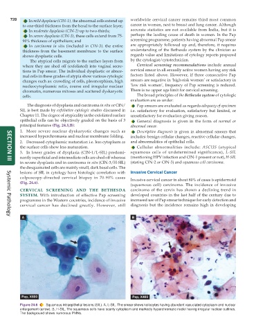
colposcopy-directed cervical biopsy in 70-90% cases Invasive cervical cancer in about 80% of cases is epidermoid
(Fig. 24.6).
(squamous cell) carcinoma. The incidence of invasive
CERVICAL SCREENING AND THE BETHESDA carcinoma of the cervix has shown a declining trend in
SYSTEM. With introduction of effective Pap screening developed countries in the last half of the century due to
programme in the Western countries, incidence of invasive increased use of Pap smear technique for early detection and
cervical cancer has declined greatly. However, still diagnosis but the incidence remains high in developing
Systemic Pathology
Figure 24.6 Squamous intraepithelial lesions (SIL). A, L-SIL. The smear shows koilocytes having abundant vacuolated cytoplasm and nuclear
enlargement (arrow). B, H-SIL. The squamous cells have scanty cytoplasm and markedly hyperchromatic nuclei having irregular nuclear outlines.
The background shows numerous PMNs.