
Page 760 - Textbook of Pathology, 6th Edition
P. 760
744
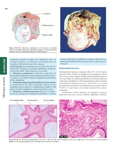
Figure 24.23 Mucinous cystadenoma of the ovary. Cut surface
shows a very large multilocular cyst without papillae. The cyst wall shows
presence of loculi containing gelatinous mucoid material.
malignant varieties usually have thickened areas. In forming solid sheets, papillary formation, adenomatous
younger patients, an element of teratoma may be pattern and infiltration into stroma with or without pools
recognised in the firm areas of the tumour. of mucin.
Histologically, the most distinctive feature is the charac-
teristic tall columnar nonciliated epithelium lining the Endometrioid Tumours
loculi (Fig. 24.24). Other features are as under: Endometrioid tumours comprise about 5% of all ovarian
1. Mucinous cystadenoma is lined by a single layer of tumours. Most of them are malignant accounting for about
SECTION III
these cells having basal nuclei and apical mucinous vacuo- 20% of all ovarian cancers. Benign and borderline forms are
les. There is very little tendency to papillary proliferation very rare. They are called endometrioid carcinomas because
of the epithelium. of the close resemblance of histologic pattern to that of uterine
2. Borderline (atypical proliferating) mucinous tumour endometrioid adenocarcinoma. About 40% of ovarian
is identified by the same histologic criteria as for endometrioid carcinomas have bilateral involvement. About
borderline serous tumour i.e. stratification (usually 2-3 cell 15-30% of cases have coexistent endometrial adeno-
thick) of typical epithelium without stromal invasion. carcinoma.
3. Mucinous cystadenocarcinoma likewise is charac- Histogenesis of these tumours in majority of cases is
terised by piling up of malignant epithelium, at places
believed to be from ovarian coelomic epithelium differen-
Systemic Pathology
Figure 24.24 Mucinous cystadenoma of the ovary. The cyst wall and the septa are lined by a single layer of tall columnar mucin-secreting
epithelium with basally-placed nuclei and large apical mucinous vacuoles.