
Page 781 - Textbook of Pathology, 6th Edition
P. 781
epidermal cells, spherical, having hyperchromatic nuclei 765
with cytoplasmic halo that stains positively with
mucicarmine. In these respects, Paget’s cells are
adenocarcinoma-type cells. In addition, the underlying
breast contains invasive or non-invasive duct carcinoma
which shows no obvious direct invasion of the skin of
nipple.
GRADING, STAGING AND PROGNOSIS
Histologic grading and clinical staging of breast cancer
determines the management and clinical course in these
patients.
A. HISTOLOGIC GRADING. The breast cancers are
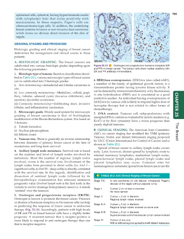
subdivided into various histologic grades depending upon Figure 25.14 Oestrogen and progesterone hormonal receptors (ER
the following parameters: and PR) in breast cancer. The tumour cells show nuclear positivity with
ER and PR antibody immunostains.
1. Histologic type of tumour. Based on classification descri-
bed in Table 25.1, various microscopic types of breast cancer
can be subdivided into 3 histologic grades: 6. HER2/neu overexpression. HER2/neu (also called erbB2),
a member of the family of epidermal growth factors, is a
i) Non-metastasising—Intraductal and lobular carcinoma in transmembrane protein having tyrosine kinase activity. It
situ.
ii) Less commonly metastasising—Medullary, colloid, papi- can be detected by immunohistochemistry or by fluorescence
llary, tubular, adenoid cystic (invasive cribriform), and in situ hybridisation (FISH) and is considered as a good
secretory (juvenile) carcinomas. predictive marker. An individual having overexpression of
iii) Commonly metastasising—Infiltrating duct, invasive HER2/neu by tumour cells is likely to respond higher dose of CHAPTER 25
herceptin therapy but is not related to other forms of
lobular, and inflammatory carcinomas.
chemotherapy.
2. Microscopic grade. Widely used system for microscopic 7. DNA content. Tumour cell subpopulations with
grading of breast carcinoma is that of Nottingham aneuploid DNA content as evaluated by mitotic markers (e.g.
modification of the Bloom-Richardson system. It is based on Ki-67) or by flow cytometry have a worse prognosis than
3 features: purely diploid tumours.
i) Tubule formation
ii) Nuclear pleomorphism B. CLINICAL STAGING. The American Joint Committee The Breast
iii) Mitotic count. (AJC) on cancer staging has modified the TNM (primary
Tumour, Nodal, and distant Metastasis) staging proposed
3. Tumour size. There is generally an inverse relationship by UICC (Union International for Control of Cancer) and is
between diameter of primary breast cancer at the time of shown in Table 25.2.
mastectomy and long-term survival.
Spread of breast cancer to axillary lymph nodes occurs
4. Axillary lymph node metastasis. Survival rate is based early. Later, however, distant spread by lymphatic route to
on the number and level of lymph nodes involved by internal mammary lymphatics, mediastinal lymph nodes,
metastasis. More the number of regional lymph nodes supraclavicular lymph nodes, pleural lymph nodes and
involved, worse is the survival rate. Involvement of the pleural lymphatics may occur. Common sites for
lymph nodes from proximal to distal axilla (i.e. level I— haematogenous metastatic spread from breast cancer are the
superficial axilla, to level III—deep axilla) is directly correlated
with the survival rate. In this regards, identification and
dissection of sentinel lymph node followed by its TABLE 25.2: AJC Clinical Staging of Breast Cancer.
histopathologic examination has attained immense Stage TIS: In situ carcinoma (in situ lobular, intraductal, Paget’s
prognostic value (Sentinel lymph node is the first node in the disease of the nipple without palpable lump)
vicinity to receive drainage from primary cancer i.e. it stands Stage I: Tumour 2 cm or less in diameter
‘sentinel’ over the tumour). No nodal spread
5. Oestrogen and progesterone receptors (ER/PR). Stage II: Tumour > 2 cm in diameter
Oestrogen is known to promote the breast cancer. Presence Regional lymph nodes involved
or absence of hormone receptors on the tumour cells can help
in predicting the response of breast cancer to endocrine Stage III A: Tumour > 5 cm in diameter
Regional lymph nodes involved on same side
therapy (Fig. 25.14). Accordingly, patients with high levels
of ER and PR on breast tumour cells have a slightly better Stage III B: Tumour > 5 cm in diameter
prognosis. A recurrent tumour that is receptor-positive is Supraclavicular and infraclavicular lymph nodes involved
more likely to respond to anti-oestrogen therapy than one Stage IV: Tumour of any size
that is receptor-negative. With or without regional spread but with distant metastasis