
Page 780 - Textbook of Pathology, 6th Edition
P. 780
764 5. METAPLASTIC CARCINOMA. Rarely, invasive
ductal carcinomas may have various types of metaplastic
alterations such as squamous metaplasia, cartilagenous and
osseous metaplasia, or their combinations. Development of
squamous cell carcinoma of the breast parenchyma is
exceedingly rare and must be separated from lesions of
epidermis or nipple region.
C. PAGET’S DISEASE OF THE NIPPLE
Paget’s disease of the nipple is an eczematoid lesion of the
nipple, often associated with an invasive or non-invasive
ductal carcinoma of the underlying breast. The nipple bears
a crusted, scaly and eczematoid lesion with a palpable
subareolar mass in about half the cases. Most of the patients
with palpable mass are found to have infiltrating duct
carcinoma, while those with no palpable breast lump are
usually subsequently found to have intraductal carcinoma.
Prognosis of patients with ductal carcinoma having Paget’s
disease is less favourable than of those who have ductal
carcinoma without Paget’s disease.
Figure 25.12 Colloid (mucinous) carcinoma breast. The tumour cells
are seen as clusters floating in pools of abundant mucin. The pathogenesis of Paget’s disease of the breast is
explained by the following 2 hypotheses:
3. SECRETORY (JUVENILE) CARCINOMA. This pattern 1. The tumour cells from the underlying ductal carcinoma
is found more frequently in children and has a better have migrated up into the lactiferous ducts and invaded the
prognosis. The tumour is generally circumscribed which epidermis producing skin lesions.
on histologic examination shows abundant intra- and 2. An alternate theory, though less reliable than the former,
extracellular PAS-positive clear spaces due to secretory is that Paget’s disease represents a form of carcinoma in situ
activity of tumour cells. of the epidermis itself.
SECTION III
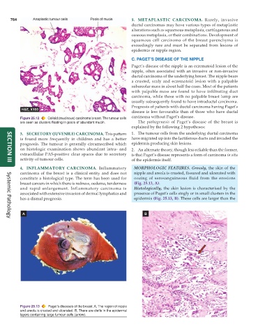
4. INFLAMMATORY CARCINOMA. Inflammatory MORPHOLOGIC FEATURES. Grossly, the skin of the
carcinoma of the breast is a clinical entity and does not nipple and areola is crusted, fissured and ulcerated with
constitute a histological type. The term has been used for oozing of serosanguineous fluid from the erosions
breast cancers in which there is redness, oedema, tenderness (Fig. 25.13, A).
and rapid enlargement. Inflammatory carcinoma is Histologically, the skin lesion is characterised by the
associated with extensive invasion of dermal lymphatics and presence of Paget’s cells singly or in small clusters in the
has a dismal prognosis. epidermis (Fig. 25.13, B). These cells are larger than the
Systemic Pathology
Figure 25.13 Paget’s diseases of the breast. A, The region of nipple
and areola is crusted and ulcerated. B, There are clefts in the epidermal
layers containing large tumour cells (arrow).