
Page 782 - Textbook of Pathology, 6th Edition
P. 782
766
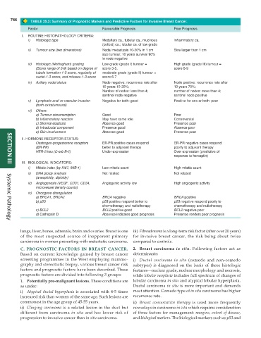
TABLE 25.3: Summary of Prognostic Markers and Predictive Factors for Invasive Breast Cancer.
Factor Favourable Prognosis Poor Prognosis
I. ROUTINE HISTOPATHOLOGY CRITERIA:
i) Histologic type Medullary ca., tubular ca., mucinous Inflammatory ca.
(colloid) ca.; lobular ca. of low grade
ii) Tumour size (two dimensions) Nodal metastasis 10-20% in 1 cm Size larger than 1 cm
size tumour; 10 years survival 90%
in node negative
iii) Histologic (Nottingham) grading Low grade (grade I) tumour = High grade (grade III) tumour =
(Score range of 3-9) based on degree of score 3-5, score 8-9
tubule formation-1-3 score, regularity of moderate grade (grade II) tumour =
nuclei-1-3 score, and mitoses-1-3 score score 6-7
iv) Axillary nodal status Node negative: recurrence rate after Node positive: recurrence rate after
10 years 10-30%; 10 years 70%;
Number of nodes: less than 4; number of nodes: more than 4;
sentinel node negative sentinel node positive
v) Lymphatic and/ or vascular invasion Negative for both: good Positive for one or both: poor
(both extratumoural)
vi) Others:
a) Tumour circumscription Good Poor
b) Inflammatory reaction May have some role Controversial
c) Stromal elastosis Absence good Presence poor
d) Intraductal component Presence good Absence poor
e) Skin involvement Absence good Presence poor
II. HORMONE RECEPTOR STATUS:
Oestrogen-progesterone receptors ER-PR positive cases respond ER-PR negative cases respond
(ER-PR) better to adjuvant therapy poorly to adjuvant therapy
HER-2/neu (C-erb B-2) Under expression Over expression (predictive of
response to herceptin)
III. BIOLOGICAL INDICATORS:
SECTION III
i) Mitotic index (by Ki67, MIB-1) Low mitotic count High mitotic count
ii) DNA ploidy analysis Not related Not related
(aneuploidy, diploidy)
iii) Angiogenesis (VEGF, CD31, CD34, Angiogenic activity low High angiogenic activity
microvessel density counts)
iv) Oncogene disregulation
a) BRCA1, BRCA2 BRCA negative BRCA positive
b) p53 p53 positive respond better to p53 negative respond poorly to
chemotherapy and radiotherapy chemotherapy and radiotherapy
c) BCL2 BCL2 positive good BCL2 negative poor
d) Cathepsin D Absence indicates good prognosis Presence renders poor prognosis
Systemic Pathology
lungs, liver, bones, adrenals, brain and ovaries. Breast is one iii) Fibroadenoma is a long-term risk factor (after over 20 years)
of the most suspected source of inapparent primary for invasive breast cancer, the risk being about twice
carcinoma in women presenting with metastatic carcinoma. compared to controls.
C. PROGNOSTIC FACTORS IN BREAST CANCER. 2. Breast carcinoma in situ. Following factors act as
Based on current knowledge gained by breast cancer determinants:
screening programmes in the West employing mammo- i) Ductal carcinoma in situ (comedo and non-comedo
graphy and stereotactic biopsy, various breast cancer risk subtypes) is diagnosed on the basis of three histologic
factors and prognostic factors have been described. These features—nuclear grade, nuclear morphology and necrosis,
prognostic factors are divided into following 3 groups: while lobular neoplasia includes full spectrum of changes of
1. Potentially pre-malignant lesions. These conditions are lobular carcinoma in situ and atypical lobular hyperplasia.
as under: Ductal carcinoma in situ is more important and demands
i) Atypical ductal hyperplasia is associated with 4-5 times most attention. Comedo type of in situ carcinoma has higher
increased risk than women of the same age. Such lesions are recurrence rate.
commonest in the age group of 45-55 years. ii) Breast conservative therapy is used more frequently
ii) Clinging carcinoma is a related lesion in the duct but nowadays in carcinoma in situ which requires consideration
different from carcinoma in situ and has lower risk of of three factors for management: margins, extent of disease,
progression to invasive cancer than in situ carcinoma. and biological markers. The biological markers such as p53 and