
Page 784 - Textbook of Pathology, 6th Edition
P. 784
768
Chapter 26 The Skin
Chapter 26
NORMAL STRUCTURE
The skin or the integument is the external organ that protects
against mechanical trauma, UV light and infection. In
addition, the skin is concerned with thermoregulation,
conservation and excretion of fluid, sensory perception and,
of course, has aesthetic role for appearance of the indidivdual.
The histology of normal skin shows some variation in
different parts of the body. In general, it is composed of 2
layers, the epidermis and the dermis, which are separated
by an irregular border. Cone-shaped dermal papillae extend
upward into the epidermis forming peg-like rete ridges of the
epidermis. Fig. 26.1 presents a diagrammatic representation
of the main structures identifiable in a section of the normal
skin while Fig. 26.2 shows the various layers of the epidermis.
EPIDERMIS
The epidermis is composed of the following 5 layers from
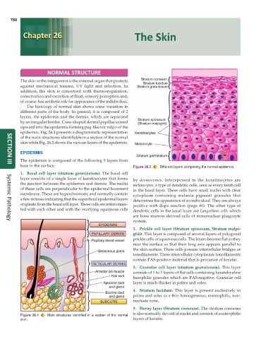
base to the surface: Figure 26.2 Different layers comprising the normal epidermis.
SECTION III
1. Basal cell layer (stratum germinatum). The basal cell
layer consists of a single layer of keratinocytes that forms by desmosomes. Interspersed in the keratinocytes are
the junction between the epidermis and dermis. The nuclei melanocytes, a type of dendritic cells, seen as every tenth cell
of these cells are perpendicular to the epidermal basement in the basal layer. These cells have small nuclei with clear
membrane. These are hyperchromatic and normally contain cytoplasm containing melanin pigment granules that
a few mitoses indicating that the superficial epidermal layers determines the appearance of an individual. They are always
originate from the basal cell layer. These cells are interconnec- positive with dopa reaction (page 40). The other type of
ted with each other and with the overlying squamous cells dendritic cells in the basal layer are Langerhans cells which
are bone marrow-derived cells of mononuclear-phagocyte
system.
Systemic Pathology
2. Prickle cell layer (Stratum spinosum, Stratum malpi-
ghii). This layer is composed of several layers of polygonal
prickle cells or squamous cells. The layers become flat as they
near the surface so that their long axis appears parallel to
the skin surface. These cells possess intercellular bridges or
tonofilaments. These intercellular cytoplasmic tonofilaments
contain PAS-positive material that is precursor of keratin.
3. Granular cell layer (stratum granulosum). This layer
consists of 1 to 3 layers of flat cells containing keratohyaline
basophilic granules which are PAS-negative. Granular cell
layer is much thicker in palms and soles.
4. Stratum lucidum. This layer is present exclusively in
palms and soles as a thin homogeneous, eosinophilic, non-
nucleate zone.
5. Horny layer (Stratum corneum). The stratum corneum
is also normally devoid of nuclei and consists of eosinophilic
Figure 26.1 Main structures identified in a section of the normal
skin. layers of keratin.