
Page 789 - Textbook of Pathology, 6th Edition
P. 789
nerves and the tissues supplied by the nerves. The condition 773
is characterised by sharp burning pain, often dispropor-
tionate to the rash. Herpes simplex, caused by HSV-1, and
another related herpetic infection, herpes genitalis, caused
by HSV-2, are characterised by transmission by direct
physical contact and prolonged latency. The vesicular lesions
are often located on the skin, especially the facial skin around
lips and external nares; other sites are mucosal surfaces and
eyes.
Histologically, the characteristic feature of viral exan-
themata is the formation of intra-epidermal vesicles or
bullae due to cytopathic effects of viruses. In the early
stage, there is proliferation of epidermal cells and
formation of multinucleate giant cells. This is followed
by intracellular oedema and ballooning degeneration that
progresses on to rupture of the cells with eventual
formation of vesicles or bullae.
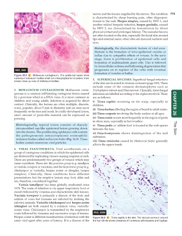
Figure 26.5 Molluscum contagiosum. The epidermal layers show
numerous molluscum bodies which are intracytoplasmic inclusions. Inset 5. SUPERFICIAL MYCOSES. Superficial fungal infections
shows close-up view of molluscum bodies.
of the skin are localised to stratum corneum (page 183). These
include some of the common dermatophytes such as
3. MOLLUSCUM CONTAGIOSUM. Molluscum conta- Trichophyton rubrum and Pityrosporum. Clinically, these fungal
giosum is a common self-limiting contagious lesion caused infections are labelled according to the region involved. These
by a poxvirus which is a DNA virus. It is more common in are as follows:
children and young adults. Infection is acquired by direct i) Tinea capitis occurring on the scalp, especially in
contact. Clinically, the lesions are often multiple, discrete, children. CHAPTER 26
waxy, papules, about 5 mm in diameter and are seen more ii) Tinea barbae affecting the region of beard in adult males.
frequently on the face and trunk. In a fully-developed lesion,
small amount of paste-like material can be expressed on iii) Tinea corporis involving the body surface at all ages.
pressing. iv) Tinea cruris occurs most frequently in the region of groin
in obese men, especially in hot weather.
Histologically, typical lesion consists of sharply v) Tinea pedis or ‘athlete foot’ is located in the web spaces
circumscribed cup-like epidermal lesion growing down between the toes. The Skin
into the dermis. The proliferating epidermal cells contain vi) Onychomycosis shows disintegration of the nail
the pathognomonic intracytoplasmic eosinophilic substance.
inclusion bodies called molluscum bodies (Fig. 26.5). These vii) Tinea versicolor caused by Malassezia furfur generally
bodies contain numerous viral particles.
affects the upper trunk.
4. VIRAL EXANTHEMATA. Viral exanthemata are a
group of contagious conditions in which the epidermal cells
are destroyed by replicating viruses causing eruption or rash.
There are predominantly two groups of viruses which may
cause exanthem. These are: the poxvirus group (e.g. smallpox
or variola, cowpox or vaccinia), and the herpesvirus group (e.g.
chickenpox or varicella, herpes zoster or shingles, herpes
simplex). Clinically, these conditions have different
presentations but the eruptive lesions may look alike and
are, therefore, considered together.
Variola (smallpox) has been globally eradicated since
1978. The route of infection is via upper respiratory tract or
mouth followed by viraemia and characteristic skin lesions.
Vaccinia (cowpox) is primarily a disease of the teats and
udders of cows but humans are infected by milking the
infected animals. Varicella (chickenpox) and herpes zoster
(shingles) are both caused by a common virus, varicella-
zoster virus. Chickenpox is transmitted by the respiratory
route followed by viraemia and successive crops of lesions.
Herpes zoster is different manifestation of infection with the Figure 26.6 Tinea capitis in the skin. The stratum corneum around
same viral agent after years of latency. It is a disease of the the hair follicle shows presence of numerous arthrospores and hyphae.