
Page 793 - Textbook of Pathology, 6th Edition
P. 793
epidermal regeneration from the periphery at the floor of 777
the bulla. The bullous cavity contains fibrin network and
many mononuclear inflammatory cells and many
eosinophils (Fig. 26.12). Dermal changes seen in
inflammatory bullae consist of infiltrate of mononuclear
cells, a few eosinophils and neutrophils.
3. DERMATITIS HERPETIFORMIS. Dermatitis herpeti-
formis is a form of chronic, pruritic, vesicular dermatosis.
The lesions are found more commonly in males in 3rd to 4th
decades of life. The disease has an association with gluten-
sensitive enteropathy (coeliac disease). Both dermatitis
herpetiformis and gluten-sensitive enteropathy respond to
a gluten-free diet. The pathogenesis of the disease is not quite
clear but probably individuals with certain histocompatibility
types develop IgA and IgG antibodies to gliadin which is a
fraction of gluten present in the flour (page 575).
Histologically, the early lesions of dermatitis herpeti-
formis consist of neutrophilic micro-abscesses at the tips
of papillae, producing separation or blister between the
papillary dermis and the epidermis (Fig. 26.13). The older
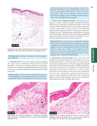
Figure 26.12 Bullous pemphigoid. The skin shows non-acantholytic blisters contain fair number of eosinophils causing
subepidermal bulla containing microabscess of eosinophils (arrow). confusion with bullous pemphigoid. Direct immuno-
fluorescence shows granular deposits of IgA at the
papillary tips in dermatitis herpetiformis.
Histologically, the picture is identical to that of pemphi- CHAPTER 26
gus foliaceous. 4. ERYTHEMA MULTIFORME. This is an acute, self-
limiting but recurrent dermatosis. The condition occurs due
2. PEMPHIGOID. This is a form of bullous disease affecting to hypersensitivity to certain infections and drugs, and in
skin or the mucous membranes. Three variants have been many cases, it is idiopathic. As the name suggests, the lesions
described—localised form occurring on the lower extremities; are multiform such as macular, papular, vesicular and bullous.
vesicular form consisting of small tense blisters; and vegetating Quite often, the lesions have symmetric involvement of the
form having verrucous vegetations found mainly in the axillae extremities. Stevens-Johnson syndrome is a severe, at times The Skin
and groins. fatal, form of involvement of skin and mucous membranes
of the mouth, conjunctivae, genital and perianal area.
Histologically, the characteristic distinguishing feature Another variant termed toxic epidermal necrolysis consists of
from pemphigus is the subepidermal location of the non- diffuse necrosis of the epidermis and mucosa, exposing the
acantholytic bullae. With passage of time, there is some dermis giving the skin a scalded appearance.
Figure 26.13 Dermatitis herpetiformis. The tips of dermal papillae Figure 26.14 Erythema multiforme. There is pronounced dermo-
show neutrophilic microabscess (arrow) causing dermo-epidermal epidermal interface dermatitis. There is oedema and necrosis of
separation at tips. kertinocytes at the junction and mild lymphocytic infiltrate.