
Page 794 - Textbook of Pathology, 6th Edition
P. 794
778
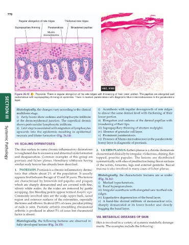
Figure 26.15 Psoriasis. There is regular elongation of the rete ridges with thickening of their lower portion. The papillae are elongated and
oedematous with suprapapillary thinning of epidermis. There is marked parakeratosis with diagnostic Munro microabscesses in the parakeratotic
layer.
Histologically, the changes vary according to the clinical i) Acanthosis with regular downgrowth of rete ridges
multiform stage. to almost the same dermal level with thickening of their
i) Early lesions show oedema and lymphocytic infiltrate lower portion.
at the dermoepidermal junction. The superfical dermis ii) Elongation and oedema of the dermal papillae with
SECTION III
shows perivascular lymphocytic infiltrate. broadening of their tips.
ii) Later stage is associated with migration of lymphocytes iii) Suprapapillary thinning of stratum malpighii.
upwards into the epidermis resulting in epidermal iv) Absence of granular cell layer.
necrosis and blister formation (Fig. 26.14). v) Prominent parakeratosis.
vi) Presence of Munro microabscesses in the parakeratotic
VII. SCALING DERMATOSES horny layer is diagnostic of psoriasis.
The skin surface in some chronic inflammatory dermatoses 2. LICHEN PLANUS. Lichen planus is a chronic dermatosis
is roughened due to excessive and abnormal scale formation characterised clinically by irregular, violaceous, shining, flat-
and desquamation. Common examples of this group are topped, pruritic papules. The lesions are distributed
psoriasis and lichen planus. Hereditary ichthyosis having symmetrically with sites of predilection being flexor surfaces
Systemic Pathology
similar scaly lesions has already been described. of the wrists, forearms, legs and external genitalia. Buccal
1. PSORIASIS. Psoriasis is a chronic inflammatory derma- mucosa is also involved in many cases of lichen planus.
tosis that affects about 2% of the population. It usually Histologically, the characteristic features are as under
appears first between the age of 15 and 30 years. The lesions (Fig. 26.16):
are characterised by brownish-red papules and plaques i) Marked hyperkeratosis.
which are sharply demarcated and are covered with fine, ii) Focal hypergranulosis.
silvery white scales. As the scales are removed by gentle iii) Irregular acanthosis with elongated saw-toothed rete
scrapping, fine bleeding points appear termed Auspitz sign. ridges.
Commonly involved sites are the scalp, upper back, sacral iv) Liquefactive degeneration of the basal layer.
region and extensor surfaces of the extremities, especially v) A band-like dermal infiltrate of mononuclear cells,
the knees and elbows. In about 25% of cases, peculiar pitting sharply demarcated at its lower border and closely
of nails is seen. Psoriatic arthritis resembling rheumatoid hugging the basal layer.
arthritis is produced in about 5% of cases but rheumatoid
factor is absent.
VIII. METABOLIC DISEASES OF SKIN
Histologically, the following features are observed in Skin is involved in a variety of systemic metabolic derange-
fully-developed lesions (Fig. 26.15):
ments. The examples include the following: