
Page 790 - Textbook of Pathology, 6th Edition
P. 790
774 Histologically, fungal hyphae (or mycelia) and
arthrospores of dermatophytes are present in the stratum
corneum of skin, nails or hair. Hyphae may be septate or
nonseptate. Spores are round to oval bodies which grow
by budding. Special stains can be used to demonstrate
the fungi. These are: periodic acid-Schiff (PAS) reaction
which stains the fungi deep pink to red (Fig. 26.6), and
methenamine silver nitrate method that stains fungi black.
IV.GRANULOMATOUS DISEASES
In many skin diseases, the host may respond by granu-
lomatous inflammation to a variety of microbial agents and
nonmicrobial material. Tuberculosis of the skin is the classical
example in which typical tubercles are formed; other
conditions are leprosy, syphilis, sarcoidosis, deep fungal
infection etc. These conditions have already been discussed
in Chapter 6. Nonmicrobial agents which can incite
granulomatous inflammation are keratin, hair, thorns, talc,
minerals like beryllium, asbestos and tattoo pigment etc.
Important representative examples of granulomatous
inflammation—lupus vulgaris, cutaneous sarcoidosis and
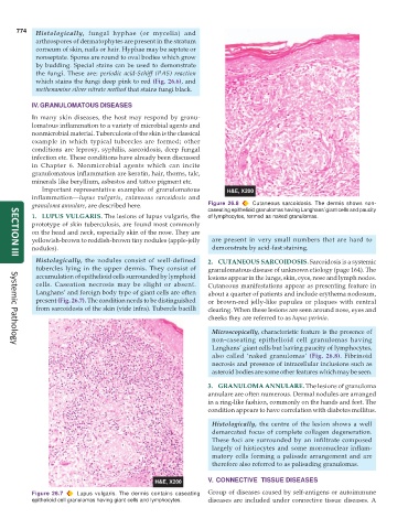
granuloma annulare, are described here. Figure 26.8 Cutaneous sarcoidosis. The dermis shows non-
caseating epithelioid granulomas having Langhans’ giant cells and paucity
1. LUPUS VULGARIS. The lesions of lupus vulgaris, the of lymphocytes, termed as naked granulomas.
prototype of skin tuberculosis, are found most commonly
on the head and neck, especially skin of the nose. They are
yellowish-brown to reddish-brown tiny nodules (apple-jelly are present in very small numbers that are hard to
nodules). demonstrate by acid-fast staining.
Histologically, the nodules consist of well-defined 2. CUTANEOUS SARCOIDOSIS. Sarcoidosis is a systemic
SECTION III
tubercles lying in the upper dermis. They consist of granulomatous disease of unknown etiology (page 164). The
accumulation of epithelioid cells surrounded by lymphoid lesions appear in the lungs, skin, eyes, nose and lymph nodes.
cells. Caseation necrosis may be slight or absent. Cutaneous manifestations appear as presenting feature in
Langhans’ and foreign body type of giant cells are often about a quarter of patients and include erythema nodosum,
present (Fig. 26.7). The condition needs to be distinguished or brown-red jelly-like papules or plaques with central
from sarcoidosis of the skin (vide infra). Tubercle bacilli clearing. When these lesions are seen around nose, eyes and
cheeks they are referred to as lupus perinio.
Microscopically, characteristic feature is the presence of
non-caseating epithelioid cell granulomas having
Langhans’ giant cells but having paucity of lymphocytes,
Systemic Pathology
also called ‘naked granulomas’ (Fig. 26.8). Fibrinoid
necrosis and presence of intracellular inclusions such as
asteroid bodies are some other features which may be seen.
3. GRANULOMA ANNULARE. The lesions of granuloma
annulare are often numerous. Dermal nodules are arranged
in a ring-like fashion, commonly on the hands and feet. The
condition appears to have correlation with diabetes mellitus.
Histologically, the centre of the lesion shows a well
demarcated focus of complete collagen degeneration.
These foci are surrounded by an infiltrate composed
largely of histiocytes and some mononuclear inflam-
matory cells forming a palisade arrangement and are
therefore also referred to as palisading granulomas.
V. CONNECTIVE TISSUE DISEASES
Figure 26.7 Lupus vulgaris. The dermis contains caseating Group of diseases caused by self-antigens or autoimmune
epithelioid cell granulomas having giant cells and lymphocytes. diseases are included under connective tissue diseases. A