
Page 792 - Textbook of Pathology, 6th Edition
P. 792
776
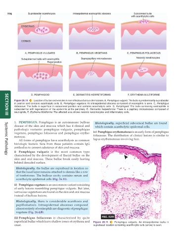
Figure 26.10 Location of bullae and vesicles in non-infectious bullous dermatoses. A, Pemphigus vulgaris: The bulla is predominantly suprabasilar
in position and contains acantholytic cells. B, Pemphigus vegetans: An intraepidermal abscess composed of eosinophils is seen. C, Pemphigus
foliaceous: The bulla is superficial in subcorneal position and contains acantholytic cells. D, Pemphigoid: The bulla containing eosinophilis is
subepidermal with regeneration of the epidermis at the periphery. E, Dermatitis herpetiformis: There is a papillary microabscess composed of
neutrophils. F, Erythema Multiforme: The affected area shows necrotic keratinocytes and inflammatory cells.
SECTION III
1. PEMPHIGUS. Pemphigus is an autoimmune bullous Histologically, superficial subcorneal bullae are found
disease of the skin and mucosa which has 4 clinical and which contain acantholytic epidermal cells.
pathologic variants: pemphigus vulgaris, pemphigus
vegetans, pemphigus foliaceous and pemphigus erythe- iv) Pemphigus erythematosus is an early form of pemphigus
matosus. foliaceous. The distribution of clinical lesions is similar to
All forms of pemphigus have acantholysis as common lupus erythematosus involving face.
histologic feature. Sera from these patients contain IgG
antibodies to cement substance of skin and mucosa.
i) Pemphigus vulgaris is the most common type
characterised by the development of flaccid bullae on the
Systemic Pathology
skin and oral mucosa. These bullae break easily leaving
behind denuded surface.
Histologically, the bullae are suprabasal in location so
that the basal layer remains attached to dermis like a row
of tombstones. The bullous cavity contains serum and
acantholytic epidermal cells (Fig. 26.11).
ii) Pemphigus vegetans is an uncommon variant consisting
of early lesions resembling pemphigus vulgaris. But later,
verrucous vegetations are found on the skin and oral mucosa
instead of bullous lesions.
Histologically, there is considerable acanthosis and
papillomatosis. Intraepidermal abscesses composed
almost entirely of eosinophils are diagnostic of pemphigus
vegetans (Fig. 24.4,B).
iii) Pemphigus foliaceous is characterised by quite
superficial bullae which leave shallow zones of erythema and Figure 26.11 Pemphigus vulgaris. An intraepidermal bulla in
crust. suprabasal location containing acantholytic cells (arrow) is seen.