
Page 791 - Textbook of Pathology, 6th Edition
P. 791
list of such diseases along with their etiology and patho- Direct immunofluorescence reveals granular deposits of 775
genesis is given in Chapter 4. Morphology of skin lesions of immunoglobulins, most commonly IgG and IgM, and
two important representative examples—lupus erythematosus components of complement on the basement membrane of
and systemic sclerosis (scleroderma), is given below. Another the affected skin in both DLE and SLE. High serum titres of
connective tissue disease of unknown etiology, lichen antinuclear antibodies and demonstration of LE cells (page
sclerosus et atrophicus, is also considered here. 79) are other notable features, especially in SLE.
1. LUPUS ERYTHEMATOSUS. Two types of lupus 2. SYSTEMIC SCLEROSIS (SCLERODERMA). Two
erythematosus are recognised—a chronic form, discoid lupus types of systemic sclerosis or scleroderma are identified: a
erythematosus (DLE) which is confined to the skin; and a localised form called morphea, and a generalised form called
systemic form, systemic lupus erythematosus (SLE) that has progressive systemic sclerosis. A variant of progressive
widespread visceral vascular lesions. The discoid variety is systemic sclerosis is CREST syndrome. (C = calcinosis,
more common which is generally benign, while systemic R = Raynaud’s phenomenon, E = esophageal dismotility,
form may be fatal, usually from renal involvement. The S = sclerodactyly and T = telangiectasia). Etiology and
diagnosis is made on the basis of clinical, serologic and pathogenesis of these conditions are already described
pathologic changes. The characteristic cutaneous lesions in (page 80). Morphea consists of lesions limited to the skin
DLE consist of well-defined erythematous discoid patches and subcutaneous tissue, while progressive systemic
associated with scaling and atrophy and often limited to the sclerosis consists of extensive involvement of the skin and
face. In contrast, cutaneous lesions in SLE are present only the subcutaneous tissue and has visceral lesions too. The
in a small proportion of cases and consist of erythematous, lesions generally begin in the fingers and distal extremities
slightly oedematous patches which are without significant and then extend proximally to involve the arms, shoulders,
scaling and without atrophy. neck and face.
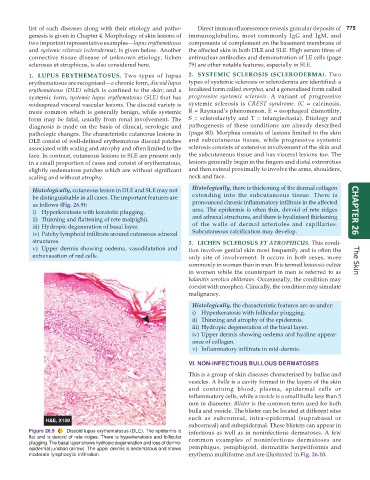
Histologically, cutaneous lesion in DLE and SLE may not Histologically, there is thickening of the dermal collagen
be distinguishable in all cases. The important features are extending into the subcutaneous tissue. There is
as follows (Fig. 26.9): pronounced chronic inflammatory infiltrate in the affected
i) Hyperkeratosis with keratotic plugging. area. The epidermis is often thin, devoid of rete ridges CHAPTER 26
ii) Thinning and flattening of rete malpighii. and adnexal structures, and there is hyalinised thickening
iii) Hydropic degeneration of basal layer. of the walls of dermal arterioles and capillaries.
iv) Patchy lymphoid infiltrate around cutaneous adnexal Subcutaneous calcification may develop.
structures. 3. LICHEN SCLEROSUS ET ATROPHICUS. This condi-
v) Upper dermis showing oedema, vasodilatation and tion involves genital skin most frequently and is often the
extravasation of red cells. only site of involvement. It occurs in both sexes, more
commonly in women than in men. It is termed kraurosis vulvae The Skin
in women while the counterpart in men is referred to as
balanitis xerotica obliterans. Occasionally, the condition may
coexist with morphea. Clinically, the condition may simulate
malignancy.
Histologically, the characteristic features are as under:
i) Hyperkeratosis with follicular plugging.
ii) Thinning and atrophy of the epidermis.
iii) Hydropic degeneration of the basal layer.
iv) Upper dermis showing oedema and hyaline appear-
ance of collagen.
v) Inflammatory infiltrate in mid-dermis.
VI. NON-INFECTIOUS BULLOUS DERMATOSES
This is a group of skin diseases characterised by bullae and
vesicles. A bulla is a cavity formed in the layers of the skin
and containing blood, plasma, epidermal cells or
inflammatory cells, while a vesicle is a small bulla less than 5
mm in diameter. Blister is the common term used for both
bulla and vesicle. The blister can be located at different sites
such as subcorneal, intra-epidermal (suprabasal or
subcorneal) and subepidermal. These blisters can appear in
Figure 26.9 Discoid lupus erythematosus (DLE). The epidermis is infectious as well as in noninfectious dermatoses. A few
flat and is devoid of rete ridges. There is hyperkeratosis and follicular common examples of noninfectious dermatoses are
plugging. The basal layer shows hydropic degeneration and loss of dermo-
epidermal junction (arrow). The upper dermis is oedematous and shows pemphigus, pemphigoid, dermatitis herpetiformis and
moderate lymphocytic infiltration. erythema multiforme and are illustrated in Fig. 26.10.