
Page 795 - Textbook of Pathology, 6th Edition
P. 795
779
Figure 26.16 Lichen planus. There is hyperkeratosis, focal hypergranulosis and irregular acanthosis with elongated saw-toothed rete ridges.
The basal layer shows liquefactive degeneration. The upper dermis shows a band-like mononuclear infiltrate with a sharply-demarcated lower
border.
1. Amyloidosis (primary as well as secondary, page 82). Many of these conditions have been discussed elsewhere CHAPTER 26
2. Lipoid proteinosis is rare. in the book; calcinosis cutis is briefly considered below.
3. Porphyria of various types (page 42). CALCINOSIS CUTIS. There are four types of calcification
4. Calcinosis cutis in the skin:
5. Gout due to urate deposits or tophi (page 853). i) Metastatic calcinosis cutis
6. Ochronosis due to alkaptonuria (page 40). ii) Dystrophic calcinosis cutis
7. Mucinosis seen in myxoedema (page 102). iii) Idiopathic calcinosis cutis The Skin
8. Idiopathic haemochromatosis with skin pigmentation iv) Subepidermal calcified nodule
(page 41).
i) Metastatic calcinosis cutis develops due to hypercal-
caemia or hyperphosphataemia as discussed on page 53.
ii) Dystrophic calcinosis cutis results when there is
deposition of calcium salts at damaged tissue.
iii) Idiopathic calcinosis cutis resembles dystrophic type but
is not associated with any underlying disease. A special
manifestation of idiopathic calcinosis cutis is tumoral calcinosis
in which there are large subcutaneous calcified masses, often
accompanied by foreign body giant cell reaction. Calcium
may discharge from the surface of the lesion. Idiopathic
calcinosis of the scrotum consists of multiple asymptomatic
nodules of the scrotal skin (Fig. 26.17).
iv) Subepidermal calcified nodule or cutaneous calculus is a
single raised hard calcified nodule in the upper dermis.
TUMOURS AND TUMOUR-LIKE LESIONS
The skin is the largest organ of the body. Tumours and
tumour-like lesions may arise from different components of
the skin such as surface epidermis, epidermal appendages
and dermal tissues. Each of these tissues may give rise to
benign and malignant tumours as well as tumour-like lesions.
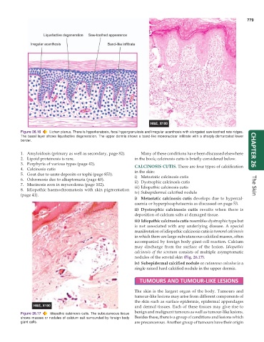
Figure 26.17 Idiopathic calcinosis cutis. The subcutaneous tissue
shows masses or nodules of calcium salt surrounded by foreign body Besides these, there is a group of conditions and lesions which
giant cells. are precancerous. Another group of tumours have their origin