
Page 798 - Textbook of Pathology, 6th Edition
P. 798
782 in sun-exposed areas of the skin in fair-skinned elderly
people. Similar lesions may be induced by exposure to
ionising radiation, hydrocarbons and arsenicals. The
condition is considered to be a forerunner of invasive
squamous cell and/or basal cell carcinoma. Clinically, the
lesions are tan-brown, erythematous, about 1 cm in diameter
with rough, sandpaper-like surface and are seen more
commonly on the dorsum of the hands and on the balded
portion of the skin.
Histologically, solar keratoses are squamous cell
carcinoma in situ with the following characteristic features:
i) Considerable hyperkeratosis.
ii) Marked acanthosis.
iii) Dyskeratosis and dysplasia of the epidermal cells
showing features such as hyperchromatism, loss of
polarity, pleomorphism and increased number of mitotic
figures.
iv) Non-specific chronic inflammatory cell infiltrate in the
upper dermis encroaching upon the basement membrane
of the epidermis.
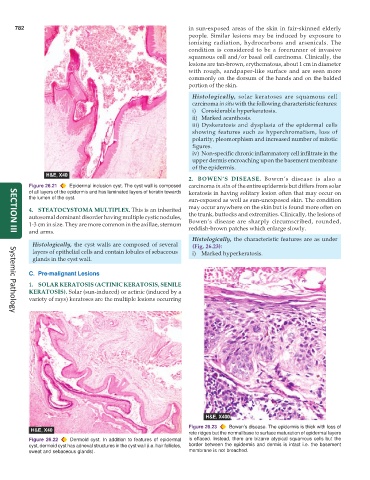
2. BOWEN’S DISEASE. Bowen’s disease is also a
Figure 26.21 Epidermal inclusion cyst. The cyst wall is composed carcinoma in situ of the entire epidermis but differs from solar
of all layers of the epidermis and has laminated layers of keratin towards keratosis in having solitary lesion often that may occur on
the lumen of the cyst. sun-exposed as well as sun-unexposed skin. The condition
4. STEATOCYSTOMA MULTIPLEX. This is an inherited may occur anywhere on the skin but is found more often on
autosomal dominant disorder having multiple cystic nodules, the trunk, buttocks and extremities. Clinically, the lesions of
1-3 cm in size. They are more common in the axillae, sternum Bowen’s disease are sharply circumscribed, rounded,
and arms. reddish-brown patches which enlarge slowly.
SECTION III
Histologically, the characteristic features are as under
Histologically, the cyst walls are composed of several (Fig. 26.23):
layers of epithelial cells and contain lobules of sebaceous i) Marked hyperkeratosis.
glands in the cyst wall.
C. Pre-malignant Lesions
1. SOLAR KERATOSIS (ACTINIC KERATOSIS, SENILE
KERATOSIS). Solar (sun-induced) or actinic (induced by a
variety of rays) keratoses are the multiple lesions occurring
Systemic Pathology
Figure 26.23 Bowen’s disease. The epidermis is thick with loss of
rete ridges but the normal base to surface maturation of epidermal layers
Figure 26.22 Dermoid cyst. In addition to features of epidermal is effaced. Instead, there are bizarre atypical squamous cells but the
cyst, dermoid cyst has adnexal structures in the cyst wall (i.e. hair follicles, border between the epidermis and dermis is intact i.e. the basement
sweat and sebaceous glands). membrane is not breached.