
Page 826 - Textbook of Pathology, 6th Edition
P. 826
810
TABLE 27.2: Contrasting Features of Simple and Nodular Goitre.
Feature Diffuse Goitre Nodular Goitre
1. Nomenclature Simple goitre, hyperplastic goitre, nontoxic goitre Multinodular, adenomatous goitre
2. Etiology Graves' disease, thyroiditis, puberty Endemic thyroiditis, cancer
3. Pathogenesis Hyperplasia-involution Repeated cycles of hyperplasia with growth and
involution with fibrosis
4. Composition Cellular-rich Colloid-rich
5. Gross Moderate, symmetric, diffuse enlargement, Nodular asymmetric, haemorrhages, scarring, cystic
colloid-filled follicles, gelatinous change, calcification
6. Microscopy Hyperplastic phase: papillary infoldings, Incomplete encapsulation, nodularity, variable-sized
Involution stage: large colloid filled follicles, fibrous scarring, haemorrhages, calcification,
follicles with flat epithelium cyst formation
7. Functional status Hyperthyroidism, euthyroid Hypothyroidism, euthyroid
The contrasting features of diffuse and nodular goitre epithelial cells forming follicles of various sizes or may
are summarised in Table 27.2. show trabecular, solid and cord patterns with little follicle
formation. Accordingly, the following 6 types of growth
THYROID TUMOURS patterns are distinguished, though more than one pattern
Most primary tumours of the thyroid are of follicular may be present in a single tumour:
epithelial origin; a few arise from parafollicular C-cells. The 1. Microfollicular (foetal) adenoma consists of small follicles
most common benign thyroid neoplasm is a follicular containing little or no colloid and separated by abundant
adenoma. Malignant tumours of the thyroid are less common loose stroma (Fig. 27.15).
but thyroid carcinoma is the most common type, though 2. Normofollicular (simple) adenoma has closely packed
rarely lymphomas and sarcomas also occur. follicles like that of normal thyroid gland.
3. Macrofollicular (colloid) adenoma contains large follicles
FOLLICULAR ADENOMA of varying size and distended with colloid.
SECTION III
4. Trabecular (embryonal) adenoma resembles embryonal
Follicular adenoma is the most common benign thyroid
tumour occurring more frequently in adult women. thyroid and consists of closely packed solid or trabecular
Clinically, it appears as a solitary nodule which can be found pattern of epithelial cells with an occasional small abortive
in approximately 1% of the population. Besides the follicular follicle.
adenoma, other conditions which may produce clinically 5. Hurthle cell (oxyphilic) adenoma is an uncommon variant
apparent solitary nodule in the thyroid are a dominant composed of solid trabeculae of large cells having
nodule of nodular goitre and thyroid carcinoma. It is thus abundant granular oxyphilic cytoplasm and vesicular
important to distinguish adenomas from these two
conditions. Though most adenomas cause no clinical problem
and behave as a ‘cold nodule’, rarely they may produce mild
hyperthyroidism and appear as ‘hot nodule’ on RAIU
Systemic Pathology
studies. Adenoma, however, rarely ever becomes malignant.
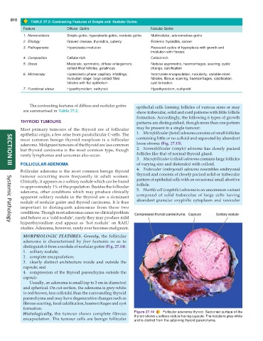
MORPHOLOGIC FEATURES. Grossly, the follicular
adenoma is characterised by four features so as to
distinguish it from a nodule of nodular goitre (Fig. 27.14):
1. solitary nodule;
2. complete encapsulation;
3. clearly distinct architecture inside and outside the
capsule; and
4. compression of the thyroid parenchyma outside the
capsule
Usually, an adenoma is small (up to 3 cm in diameter)
and spherical. On cut section, the adenoma is grey-white
to red-brown, less colloidal than the surrounding thyroid
parenchyma and may have degenerative changes such as
fibrous scarring, focal calcification, haemorrhages and cyst
formation.
Histologically, the tumour shows complete fibrous Figure 27.14 Follicular adenoma thyroid. Sectioned surface of the
encapsulation. The tumour cells are benign follicular thyroid shows a solitary nodule having capsule. The nodule is grey-white
and is distinct from the adjoining thyroid parenchyma.