
Page 825 - Textbook of Pathology, 6th Edition
P. 825
809
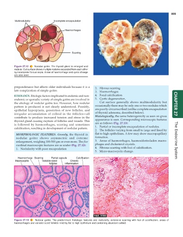
Figure 27.12 Nodular goitre. The thyroid gland is enlarged and
nodular. Cut surface shows multiple nodules separated from each other
by incomplete fibrous septa. Areas of haemorrhage and cystic change
are also seen.
preponderance but affects older individuals because it is a 2. Fibrous scarring
late complication of simple goitre. 3. Haemorrhages
ETIOLOGY. Etiologic factors implicated in endemic and non- 4. Focal calcification
endemic or sporadic variety of simple goitre are involved in 5. Cystic degeneration.
the etiology of nodular goitre too. However, how nodular Cut surface generally shows multinodularity but CHAPTER 27
pattern is produced is not clearly understood. Possibly, occasionally there may be only one or two nodules which
epithelial hyperplasia, generation of new follicles, and are poorly-circumscribed (unlike complete encapsulation
irregular accumulation of colloid in the follicles—all of thyroid adenoma, described below).
contribute to produce increased tension and stress in the Histologically, the same heterogenicity as seen on gross
thyroid gland causing rupture of follicles and vessels. This appearance is seen. Corresponding microscopic features
is followed by haemorrhages, scarring and sometimes are as follows (Fig. 27.13):
1. Partial or incomplete encapsulation of nodules.
calcification, resulting in development of nodular pattern.
2. The follicles varying from small to large and lined by
MORPHOLOGIC FEATURES. Grossly, the thyroid in flat to high epithelium. A few may show macropapillary
nodular goitre shows asymmetric and extreme formation.
enlargement, weighing 100-500 gm or even more. The five 3. Areas of haemorrhages, haemosiderin-laden macro- The Endocrine System
cardinal macroscopic features are as under (Fig. 27.12): phages and cholesterol crystals.
1. Nodularity with poor encapsulation 4. Fibrous scarring with foci of calcification.
5. Micro-macrocystic change.
Figure 27.13 Nodular goitre. The predominant histologic features are: nodularity, extensive scarring with foci of calcification, areas of
haemorrhages and variable-sized follicles lined by flat to high epithelium and containing abundant colloid.