
Page 824 - Textbook of Pathology, 6th Edition
P. 824
808
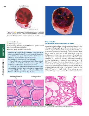
Figure 27.10 Simple (diffuse nontoxic or colloid) goitre. The thyroid
gland is enlarged diffusely. Cut section shows lobules of translucent
gelatinous light brown parenchyma and areas of haemorrhage.
Genetic factors. Nodular Goitre
Dietary goitrogenes. (Multinodular Goitre, Adenomatous Goitre)
Hereditary defect in thyroid hormone synthesis and As already stated, nodular goitre is regarded as the end-stage
transport (dyshormonogenesis). of long-standing simple goitre. It is characterised by most
Inborn errors of iodine metabolism. extreme degree of tumour-like enlargement of the thyroid
gland and characteristic nodularity. The enlargement of the
MORPHOLOGIC FEATURES. Grossly, the enlargement gland may be sufficient to not only cause cosmetic
of the thyroid gland in simple goitre is moderate (weighing disfigurement, but in many cases may cause dsyphagia and
up to 100-150 gm), symmetric and diffuse. Cut surface is choking due to compression of oesophagus and trachea. Most
SECTION III
gelatinous and translucent brown (Fig. 27.10). cases are in a euthyroid state but about 10% cases may
Histologically, two stages are distinguished: develop thyrotoxicosis resulting in toxic nodular goitre or
1. Hyperplastic stage is the early stage and is characterised
by tall columnar follicular epithelium showing papillary Plummer’s disease. However, thyrotoxicosis of Plummer’s
disease (toxic nodular goitre) differs from that of Graves’
infoldings and formation of small new follicles. disease (diffuse toxic goitre) in lacking features of ophthalmo-
2. Involution stage generally follows hyperplastic stage pathy and dermatopathy. Such ‘hot nodules’ may be picked
after variable period of time. This stage is characterised up by CT scan or by RAIU studies. Since nodular goitre is
by large follicles distended by colloid and lined by derived from simple goitre, it has the same female
flattened follicular epithelium (Fig. 27.11).
Systemic Pathology
Figure 27.11 Simple goitre. Microscopy shows large follicles distended by colloid and lined by flattened follicular epithelium.