
Page 827 - Textbook of Pathology, 6th Edition
P. 827
811
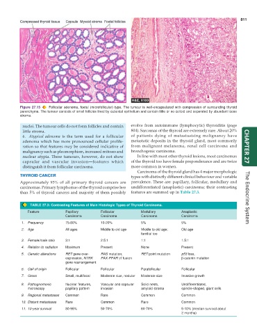
Figure 27.15 Follicular adenoma, foetal (microfollicular) type. The tumour is well-encapsulated with compression of surrounding thyroid
parenchyma. The tumour consists of small follicles lined by cuboidal epithelium and contain little or no colloid and separated by abundant loose
stroma.
nuclei. The tumour cells do not form follicles and contain evolve from autoimmune (lymphocytic) thyroiditis (page
little stroma. 804). Sarcomas of the thyroid are extremely rare. About 20%
6. Atypical adenoma is the term used for a follicular of patients dying of metastasising malignancy have
adenoma which has more pronounced cellular prolife- metastatic deposits in the thyroid gland, most commonly
ration so that features may be considered indicative of from malignant melanoma, renal cell carcinoma and CHAPTER 27
malignancy such as pleomorphism, increased mitoses and bronchogenic carcinoma.
nuclear atypia. These tumours, however, do not show In line with most other thyroid lesions, most carcinomas
capsular and vascular invasion—features which of the thyroid too have female preponderance and are twice
distinguish it from follicular carcinoma. more common in women.
Carcinoma of the thyroid gland has 4 major morphologic
THYROID CANCER types with distinctly different clinical behaviour and variable
Approximately 95% of all primary thyroid cancers are prevalence. These are: papillary, follicular, medullary and
carcinomas. Primary lymphomas of the thyroid comprise less undifferentiated (anaplastic) carcinoma; their contrasting
than 5% of thyroid cancers and majority of them possibly features are summed up in Table 27.3. The Endocrine System
TABLE 27.3: Contrasting Features of Main Histologic Types of Thyroid Carcinoma.
Feature Papillary Follicular Medullary Anaplastic
Carcinoma Carcinoma Carcinoma Carcinoma
1. Frequency 75-80% 10-20% 5% 5%
2. Age All ages Middle to old age Middle to old age; Old age
familial too
3. Female/male ratio 3:1 2.5:1 1:1 1.5:1
4. Relation to radiation Maximum Present None Present
5. Genetic alterations RET gene over- RAS mutation, RET point mutation p53 loss,
expression, NTRK PAX-PPAR γ1 fusion β-catenin mutation
gene rearrangement
6. Cell of origin Follicular Follicular Parafollicular Follicular
7. Gross Small, multifocal Moderate size, nodular Moderate size Invasive growth
8. Pathognomonic Nuclear features, Vascular and capsular Solid nests, Undifferentiated,
microscopy papillary pattern invasion amyloid stroma spindle-shaped, giant cells
9. Regional metastases Common Rare Common Common
10. Distant metastases Rare Common Rare Common
11. 10-year survival 80-95% 50-70% 60-70% 5-10% (median survival about
2 months)