
Page 861 - Textbook of Pathology, 6th Edition
P. 861
chondroid tumour with foci of haemorrhages, necrosis and 845
calcification.
Histologically, the tumour is highly cellular and is
composed of small, round to polygonal mononuclear cells
resembling chondroblasts and has multinucleate
osteoclast-like giant cells. There are small areas of
cartilaginous intercellular matrix and focal calcification.
Chondromyxoid Fibroma
Chondromyxoid fibroma is an uncommon benign tumour
of cartilaginous origin arising in the metaphysis of long
bones. Most common locations are upper end of tibia and
lower end of femur i.e. around the knee joint. Majority of
tumours appear in 2nd to 3rd decades of life with male
preponderance. Radiologically, the tumour appears as a
sharply-outlined radiolucent area with foci of calcification
and expansion of affected end of the bone. The lesion may
be asymptomatic, or may cause pain, swelling and discomfort
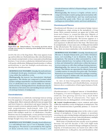
Figure 28.14 Osteochondroma. The overlying cap shows mature in the affected joint. The lesions may recur after curettage.
cartilage cells covering the underlying mature lamellar bone containing Thus, there are many similarities with chondroblastoma.
marrow spaces.
MORPHOLOGIC FEATURES. Grossly, chondromyxoid
involve the ribs or the long bones. They may appear at any fibroma is sharply-demarcated, grey-white lobulated
age and in either sex. Enchondromas, like osteochondromas, mass, not exceeding 5 cm in diameter, lying in the
may remain asymptomatic or may cause pain and pathologic metaphysis. The tumour is often surrounded by a layer
fractures. X-ray reveals a radiolucent, lobulated tumour mass of dense sclerotic bone. Cut surface of the tumour is soft CHAPTER 28
with spotty calcification. Malignant transformation of solitary to firm and lobulated but calcification within the tumour
enchondroma is rare but multiple enchondromas may is not as common as with other cartilage-forming tumours.
develop into chondrosarcoma. Histologically, the tumour has essentially lobulated
pattern. The lobules are separated by fibrous tissue and
MORPHOLOGIC FEATURES. Grossly, the enchondroma variable number of osteoclast-like giant cells. The lobules
is lobulated, bluish-grey, translucent, cartilaginous mass themselves are composed of immature cartilage consisting
lying within the medullary cavity. of spindle-shaped or stellate cells with abundant myxoid
Histologically, the tumour has characteristic lobulated or chondroid intercellular matrix.
appearance. The lobules are composed of normal adult
hyaline cartilage separated by vascularised fibrous stroma. In view of close histogenetic relationship between
Foci of calcification may be evident within the tumour. chondromyxoid fibroma and chondroblastoma, occasional
Enchondroma is distinguished from chondrosarcoma by tumours show a combination of histological features of both. The Musculoskeletal System
the absence of invasion into surrounding tissues and lack Chondrosarcoma
of cytologic features of malignancy.
Chondrosarcoma is a malignant tumour of chondroblasts.
In frequency, it is next in frequency to osteosarcoma but is
Chondroblastoma
relatively slow-growing and thus has a much better
Chondroblastoma is a relatively rare benign tumour arising prognosis than that of osteosarcoma. Two types of
from the epiphysis of long bones adjacent to the epiphyseal chondrosarcoma are distinguished: central and peripheral.
cartilage plate. Most commonly affected bones are upper tibia Central chondrosarcoma is more common and arises
and lower femur (i.e. about knee) and upper humerus. The within the medullary cavity of diaphysis or metaphysis. This
tumour usually occurs in patients under 20 years of age with type of chondrosarcoma is generally primary i.e. occurs de
male preponderance (male-female ratio 2:1). The novo.
radiographic appearance is of a sharply-circumscribed, lytic Peripheral chondrosarcoma arises in the cortex or peri-
lesion with multiple small foci of calcification. Chondro- osteum of metaphysis. It may be primary or secondary
blastoma may be asymptomatic, or may produce local pain, occurring on a pre-existing benign cartilaginous tumour such
tenderness and discomfort. The behaviour of the tumour is as osteocartilaginous exostoses (osteochondromas), multiple
benign though it may recur locally after curettage. enchondromatosis, and rarely, chondroblastoma.
Both forms of chondrosarcoma usually occur in patients
MORPHOLOGIC FEATURES. Grossly, chondroblastoma
is a well-defined mass, up to 5 cm in diameter, lying in between 3rd and 6th decades of life with slight male
the epiphysis. The tumour is surrounded by thin capsule preponderance. In contrast to benign cartilaginous tumours,
of dense sclerotic bone. Cut surface reveals a soft majority of chondrosarcomas are found more often in the
central skeleton (i.e. in the pelvis, ribs and shoulders); other