
Page 872 - Textbook of Pathology, 6th Edition
P. 872
856 Individual muscle fibre is an elongated multinucleated
syncytium-like cell about 100 μm in diameter and several
centimeters in length. The muscle nuclei are spindle-shaped
and lie at the periphery of fibre under the sarcolemma, the
plasma membrane of muscle fibre. The cytoplasm of the
muscle fibre contains myofilaments which are contractile
elements. Myofilaments are of 2 types—myosin comprising
thick filaments and actin constituting thin filaments. These
together produce cross-striations in muscle fibres seen in
longitudinal sections on light microscopy. Sarcomeres are the
partitions of myofilaments into equal zones. Each sarcomere
represents the distance between consecutive Z bands and
contains the central A (anisotropic) band, and the lateral I
(isotropic) bands.
The major functions of striated skeletal muscle are to
convert chemical energy into mechanical energy, to act as a
store of energy and proteins, and to play a role in the
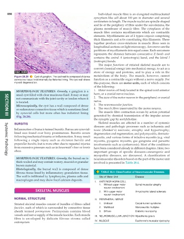
Figure 28.28 Cyst of ganglion. The cyst wall is composed of dense metabolism of the body. The muscle, however, cannot
connective tissue lined internally by flattened lining. The cyst wall shows function as a contractile organ without a nerve supply. For
myxoid degeneration. this purpose, there are motor units, each of which consists
of the following:
MORPHOLOGIC FEATURES. Grossly, a ganglion is a 1. Motor neuron cell body located in the spinal cord anterior
small cyst filled with clear mucinous fluid. It may or may horn, or a cranial nerve nucleus.
not communicate with the joint cavity or tendon where it 2. The axon of the motor neuron in the peripheral or cranial
is located. nerve.
Microscopically, the cyst has a wall composed of dense 3. The neuromuscular junction.
or oedematous connective tissue which is sometimes lined 4. The muscle fibres innervated by the motor neuron.
by synovial cells but more often has indistinct lining The muscle fibre contraction occurs by action potential
(Fig. 28.28). generated by chemical transmission of the impulse across
SECTION III
the synaptic gap by acetylcholine.
BURSITIS Skeletal muscles are affected in a number of systemic
diseases and pathologic processes such as ischaemia and
Inflammation of bursa is termed bursitis. Bursae are synovial- toxic (Zenker’s) necrosis; atrophy and hypertrophy;
lined sacs found over bony prominences. Bursitis occurs degeneration and regeneration; and polymyositis, dermato-
following mechanical trauma or inflammation. It may result myositis, and various forms of infective myositis (e.g. viral
following a single injury such as olecranon bursitis and myositis, pyogenic myositis, gas gangrene and parasitic
prepatellar bursitis, but is more often due to repeated injuries involvements such as cysticercosis). Most of the conditions
from excessive pressure such as in housemaid’s knee or tennis have been considered already in different chapters. Here, two
elbow. important groups of specific diseases—neurogenic and
myopathic diseases, are discussed. A classification of
Systemic Pathology
MORPHOLOGIC FEATURES. Grossly, the bursal sac is neuromuscular disorders based on the part of the motor unit
thick-walled and may contain watery, mucoid or granular involved is presented in Table 28.4.
brown material.
Histologically, the bursal wall is composed of dense
fibrous tissue lined by inflammatory granulation tissue. TABLE 28.4: Classification of Neuromuscular Diseases.
The wall is infiltrated by lymphocytes, plasma cells and Site of Motor Unit Disease
macrophages and may show focal calcium deposits.
I. ANTERIOR HORN CELL
1. Without upper motor Spinal muscular atrophy
neuron involvement
SKELETAL MUSCLES 2. With upper motor Amyotrophic lateral sclerosis
neuron involvement
NORMAL STRUCTURE II. PERIPHERAL NERVE
Striated skeletal muscles consist of bundles of fibres called 1. Unifocal Carpal-tunnel syndrome
fascicles, each of which is surrounded by connective tissue 2. Multifocal Mononeuritis multiplex
sheath termed perimysium. Perimysium contains blood 3. Diffuse Diabetic neuropathy
vessels and nerve supply of the muscle fascicles. Each muscle III. NEUROMUSCULAR JUNCTION Myasthenia gravis
fibre is enveloped by delicate fibrous stroma called
IV. MUSCLE Duchenne’s muscular dystrophy
endomysium.