
Page 870 - Textbook of Pathology, 6th Edition
P. 870
854 4. Renal disease involving interstitial tissue and blood therapy, drug-induced (e.g. aspirin, pyrazinamide, nicotinic
vessels. acid, ethambutol and ethanol), adrenal insufficiency,
5. Uric acid nephrolithiasis. starvation, diabetic ketosis, and disorders of parathyroid and
The disease usually begins in 3rd decade of life and affects thyroid. Renal disease per se rarely causes secondary
men more often than women. A family history of gout is hyperuricaemia such as in polycystic kidney disease and
present in a fairly large proportion of cases indicating role of leads to urate nephropathy.
inheritance in hyperuricaemia. Clinically, the natural history
of gout comprises 4 stages: asymptomatic hyperuricaemia, MORPHOLOGIC FEATURES. The pathologic mani-
acute gouty arthritis, asymptomatic intervals of intercritical festations of gout include: acute gouty arthritis, chronic
periods, and chronic tophaceous stage. In addition, gout tophaceous arthritis, tophi in soft tissues, and renal lesions
nephropathy and urate nephrolithiasis may occur (page 692). as under:
1. Acute gouty arthritis. This stage is characterised by
TYPES AND PATHOGENESIS. The fundamental bio- acute synovitis triggered by precipitation of sufficient
chemical hallmark of gout is hyperuricaemia. A serum uric amount of needle-shaped crystals of monosodium urate
acid level in excess of 7 mg/dl, which represents the upper from serum or synovial fluid. There is joint effusion
limit of solubility of monosodium urate in serum at 37°C at containing numerous polymorphs, macrophages and
blood pH, is associated with increased risk of development microcrystals of urates. The mechanism of acute
of gout. Thus, pathogenesis of gout is pathogenesis of hyper- inflammation appears to include phagocytosis of crystals
uricaemia. by leucocytes, activation of the kallikrein system, activa-
Hyperuricaemia and gout may be classified into 2 types: tion of the complement system and urate-mediated
metabolic and renal, each of which may be primary or secondary. disruption of lysosomes within the leucocytes leading to
Primary refers to cases in which the underlying biochemical release of lysosomal products in the joint effusion. Initially,
defect causing hyperuricaemia is not known, while secondary there is monoarticular involvement accompanied with
denotes cases with known causes of hyperuricaemia.
intense pain, but later it becomes polyarticular along with
1. Hyperuricaemia of metabolic origin. This group constitutional symptoms like fever. Acute gouty arthritis
comprises about 10% cases of gout which are characterised is predominantly a disease of lower extremities, affecting
by overproduction of uric acid. There is either an accelerated most commonly great toe. Other joints affected, in order
rate of purine biosynthesis de novo, or an increased turnover of decreasing frequency, are: the instep, ankles, heels,
of nucleic acids. The causes of primary metabolic gout include knees, wrists, fingers and elbows.
SECTION III
a number of specific enzyme defects in purine metabolism 2. Chronic tophaceous arthritis. Recurrent attacks of
which may be either of unknown cause or are inborn errors acute gouty arthritis lead to progressive evolution into
of metabolism. The secondary metabolic gout is due to either chronic arthritis. The deposits of urate encrust the articular
increased purine biosynthesis or a deficiency of glucose-6- cartilage. There is synovial proliferation, pannus
phosphatase. formation and progressive destruction of articular
2. Hyperuricaemia of renal origin. About 90% cases of gout cartilage and subchondral bone. Deposits of urates in the
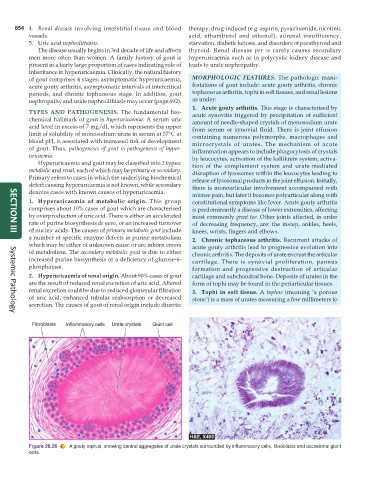
are the result of reduced renal excretion of uric acid. Altered form of tophi may be found in the periarticular tissues.
renal excretion could be due to reduced glomerular filtration 3. Tophi in soft tissue. A tophus (meaning ‘a porous
of uric acid, enhanced tubular reabsorption or decreased stone’) is a mass of urates measuring a few millimeters to
secretion. The causes of gout of renal origin include diuretic
Systemic Pathology
Figure 28.26 A gouty tophus, showing central aggregates of urate crystals surrounded by inflammatory cells, fibroblasts and occasional giant
cells.