
Page 867 - Textbook of Pathology, 6th Edition
P. 867
RA is a common disease having peak incidence in 3rd to 851
4th decades of life, with 3-5 times higher preponderance in
females. The condition has high association with HLA-DR4
and HLA-DR1 and familial aggregation. The onset of disease
is insidious, beginning with prodrome of fatigue, weakness,
joint stiffness, vague arthralgias and myalgias. This is
followed by pain and swelling of joints usually in
symmetrical fashion, especially involving joints of hands,
wrists and feet. Unlike migratory polyarthritis of rheumatic
fever, RA usually persists in the involved joint.
Approximately 20% of patients develop rheumatoid nodules
located over the extensor surfaces of the elbows and fingers.
About 80% of cases are seropositive for rheumatoid factor
(RF). However, RF titres are elevated in certain unrelated
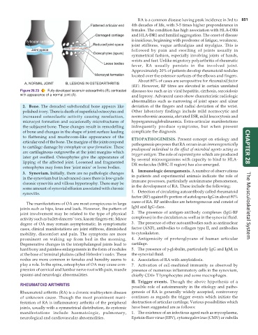
Figure 28.23 Fully-developed lesions in osteoarthritis (B), contrasted diseases too such as in: viral hepatitis, cirrhosis, sarcoidosis
with appearance of a normal joint (A). and leprosy. Advanced cases show characteristic radiologic
abnormalities such as narrowing of joint space and ulnar
2. Bone. The denuded subchondral bone appears like deviation of the fingers and radial deviation of the wrist.
polished ivory. There is death of superficial osteocytes and Other laboratory findings include mild normocytic and
increased osteoclastic activity causing rarefaction, normochromic anaemia, elevated ESR, mild leucocytosis and
microcyst formation and occasionally microfractures of hypergammaglobulinaemia. Extra-articular manifestations
the subjacent bone. These changes result in remodelling infrequently produce symptoms, but when present
of bone and changes in the shape of joint surface leading complicate the diagnosis.
to flattening and mushroom-like appearance of the ETIOPATHOGENESIS. Present concept on etiology and
articular end of the bone. The margins of the joints respond
to cartilage damage by osteophyte or spur formation. These pathogenesis proposes that RA occurs in an immunogenetically CHAPTER 28
are cartilaginous outgrowths at the joint margins which predisposed individual to the effect of microbial agents acting as
trigger antigen. The role of superantigens which are produced
later get ossified. Osteophytes give the appearance of by several microorganisms with capacity to bind to HLA-
lipping of the affected joint. Loosened and fragmented DR molecules (MHC-II region) has also emerged.
osteophytes may form free ‘joint mice’ or loose bodies.
3. Synovium. Initially, there are no pathologic changes I. Immunologic derangements. A number of observations
in the synovium but in advanced cases there is low-grade in patients and experimental animals indicate the role of
chronic synovitis and villous hypertrophy. There may be immune processes, particularly autoimmune phenomenon,
some amount of synovial effusion associated with chronic in the development of RA. These include the following:
synovitis. 1. Detection of circulating autoantibody called rheumatoid
factor (RF) against Fc portion of autologous IgG in about 80%
The manifestations of OA are most conspicuous in large cases of RA. RF antibodies are heterogeneous and consist of
joints such as hips, knee and back. However, the pattern of IgM and IgG class. The Musculoskeletal System
joint involvement may be related to the type of physical 2. The presence of antigen-antibody complexes (IgG-RF
activity such as ballet-dancers’ toes, karate fingers etc. Minor complexes) in the circulation as well as in the synovial fluid.
degree of OA may remain asymptomatic. In symptomatic 3. The presence of other autoantibodies such as antinuclear
cases, clinical manifestations are joint stiffness, diminished factor (ANF), antibodies to collagen type II, and antibodies
mobility, discomfort and pain. The symptoms are more to cytoskeleton.
prominent on waking up from bed in the morning. 4. Antigenicity of proteoglycans of human articular
Degenerative changes in the interphalangeal joints lead to cartilage.
hard bony and painless enlargements in the form of nodules 5. The presence of γ-globulin, particularly IgG and IgM, in
at the base of terminal phalanx called Heberden’s nodes. These the synovial fluid.
nodes are more common in females and heredity seems to 6. Association of RA with amyloidosis.
play a role. In the spine, osteophytes of OA may cause com- 7. Activation of cell-mediated immunity as observed by
pression of cervical and lumbar nerve root with pain, muscle presence of numerous inflammatory cells in the synovium,
spasms and neurologic abnormalities. chiefly CD4+ T lymphocytes and some macrophages.
II. Trigger events. Though the above hypothesis of a
RHEUMATOID ARTHRITIS
possible role of autoimmunity in the etiology and patho-
Rheumatoid arthritis (RA) is a chronic multisystem disease genesis of RA is generally widely accepted, controversy
of unknown cause. Though the most prominent mani- continues as regards the trigger events which initiate the
festation of RA is inflammatory arthritis of the peripheral destruction of articular cartilage. Various possibilities which
joints, usually with a symmetrical distribution, its systemic have been suggested are as follows:
manifestations include haematologic, pulmonary, 1. The existence of an infectious agent such as mycoplasma,
neurological and cardiovascular abnormalities. Epstein-Barr virus (EBV), cytomegalovirus (CMV) or rubella