
Page 871 - Textbook of Pathology, 6th Edition
P. 871
a few centimeters in diameter. Tophi may be located in 855
the periarticular tissues as well as subcutaneously such
as on the hands and feet. Tophi are surrounded by
inflammatory reaction consisting of macrophages,
lymphocytes, fibroblasts and foreign body giant cells
(Fig. 28.26).
4. Renal lesions. Chronic gouty arthritis frequently
involves the kidneys. Three types of renal lesions are
described in the kidneys: acute urate nephropathy, chronic
urate nephropathy and uric acid nephrolithiasis.
i) Acute urate nephropathy is attributed to the intratubular
deposition of monosodium urate crystals resulting in acute
obstructive uropathy.
ii) Chronic urate nephropathy refers to the deposition of
urate crystals in the renal interstitial tissue.
iii) Uric acid nephrolithiasis is related to hyperuricaemia
resulting in hyperuricaciduria (page 692).
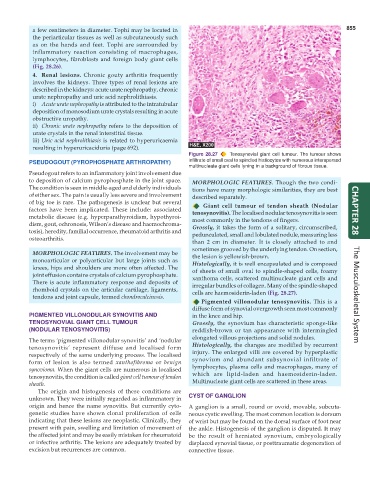
Figure 28.27 Tenosynovial giant cell tumour. The tumour shows
PSEUDOGOUT (PYROPHOSPHATE ARTHROPATHY) infiltrate of small oval to spindled histiocytes with numerous interspersed
multinucleate giant cells lyning in a background of fibrous tissue.
Pseudogout refers to an inflammatory joint involvement due
to deposition of calcium pyrophosphate in the joint space. MORPHOLOGIC FEATURES. Though the two condi-
The condition is seen in middle-aged and elderly individuals tions have many morphologic similarities, they are best
of either sex. The pain is usually less severe and involvement described separately.
of big toe is rare. The pathogenesis is unclear but several Giant cell tumour of tendon sheath (Nodular
factors have been implicated. These include: associated tenosynovitis). The localised nodular tenosynovitis is seen CHAPTER 28
metabolic disease (e.g. hyperparathyroidism, hypothyroi- most commonly in the tendons of fingers.
dism, gout, ochronosis, Wilson’s disease and haemochroma- Grossly, it takes the form of a solitary, circumscribed,
tosis), heredity, familial occurrence, rheumatoid arthritis and pedunculated, small and lobulated nodule, measuring less
osteoarthritis.
than 2 cm in diameter. It is closely attached to and
sometimes grooved by the underlying tendon. On section,
MORPHOLOGIC FEATURES. The involvement may be the lesion is yellowish-brown.
monoarticular or polyarticular but large joints such as Histologically, it is well encapsulated and is composed
knees, hips and shoulders are more often affected. The of sheets of small oval to spindle-shaped cells, foamy
joint effusion contains crystals of calcium pyrophosphate. xanthoma cells, scattered multinucleate giant cells and
There is acute inflammatory response and deposits of irregular bundles of collagen. Many of the spindle-shaped
rhomboid crystals on the articular cartilage, ligaments, cells are haemosiderin-laden (Fig. 28.27).
tendons and joint capsule, termed chondrocalcinosis. The Musculoskeletal System
Pigmented villonodular tenosynovitis. This is a
diffuse form of synovial overgrowth seen most commonly
PIGMENTED VILLONODULAR SYNOVITIS AND in the knee and hip.
TENOSYNOVIAL GIANT CELL TUMOUR Grossly, the synovium has characteristic sponge-like
(NODULAR TENOSYNOVITIS) reddish-brown or tan appearance with intermingled
elongated villous projections and solid nodules.
The terms ‘pigmented villonodular synovitis’ and ‘nodular
tenosynovitis’ represent diffuse and localised form Histologically, the changes are modified by recurrent
respectively of the same underlying process. The localised injury. The enlarged villi are covered by hyperplastic
synovium and abundant subsynovial infiltrate of
form of lesion is also termed xanthofibroma or benign lymphocytes, plasma cells and macrophages, many of
synovioma. When the giant cells are numerous in localised which are lipid-laden and haemosiderin-laden.
tenosynovitis, the condition is called giant cell tumour of tendon Multinucleate giant cells are scattered in these areas.
sheath.
The origin and histogenesis of these conditions are
unknown. They were initially regarded as inflammatory in CYST OF GANGLION
origin and hence the name synovitis. But currently cyto- A ganglion is a small, round or ovoid, movable, subcuta-
genetic studies have shown clonal proliferation of cells neous cystic swelling. The most common location is dorsum
indicating that these lesions are neoplastic. Clinically, they of wrist but may be found on the dorsal surface of foot near
present with pain, swelling and limitation of movement of the ankle. Histogenesis of the ganglion is disputed. It may
the affected joint and may be easily mistaken for rheumatoid be the result of herniated synovium, embryologically
or infective arthritis. The lesions are adequately treated by displaced synovial tissue, or posttraumatic degeneration of
excision but recurrences are common. connective tissue.