
Page 343 - First Aid for the USMLE Step 1 2020, Thirtieth edition [MedicalBooksVN.com]_Neat
P. 343
CARDIOvASCuLAR ``CARdIOvASCulAR—PATHOlOGY CARDIOvASCuLAR ``CARdIOvASCulAR—PATHOlOGY SECTION III 299
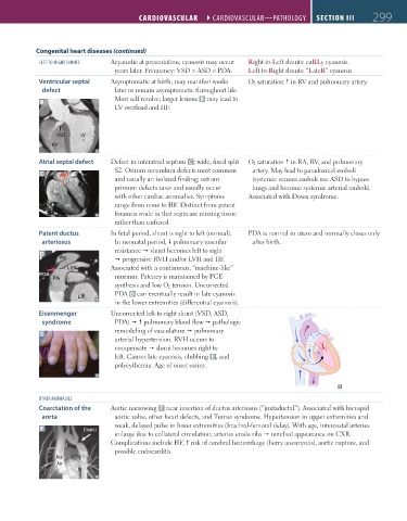
Congenital heart diseases (continued)
lEFT-TO-RIGHT SHuNTS Acyanotic at presentation; cyanosis may occur Right-to-Left shunts: eaRLy cyanosis.
years later. Frequency: VSD > ASD > PDA. Left-to-Right shunts: “LateR” cyanosis.
Ventricular septal Asymptomatic at birth, may manifest weeks O 2 saturation in RV and pulmonary artery.
defect later or remain asymptomatic throughout life.
Most self resolve; larger lesions B may lead to
B
LV overload and HF.
VSD LV
RV
Atrial septal defect Defect in interatrial septum C ; wide, fixed split O 2 saturation in RA, RV, and pulmonary
S2. Ostium secundum defects most common artery. May lead to paradoxical emboli
C ASD
and usually an isolated finding; ostium (systemic venous emboli use ASD to bypass
primum defects rarer and usually occur lungs and become systemic arterial emboli).
with other cardiac anomalies. Symptoms Associated with Down syndrome.
range from none to HF. Distinct from patent
foramen ovale in that septa are missing tissue
rather than unfused.
Patent ductus In fetal period, shunt is right to left (normal). PDA is normal in utero and normally closes only
arteriosus In neonatal period, pulmonary vascular after birth.
resistance shunt becomes left to right
D
progressive RVH and/or LVH and HF.
Associated with a continuous, “machine-like”
murmur. Patency is maintained by PGE
synthesis and low O 2 tension. Uncorrected
PDA D can eventually result in late cyanosis
in the lower extremities (differential cyanosis).
Eisenmenger Uncorrected left-to-right shunt (VSD, ASD,
syndrome PDA) pulmonary blood flow pathologic
remodeling of vasculature pulmonary
E
arterial hypertension. RVH occurs to
compensate shunt becomes right to
left. Causes late cyanosis, clubbing E , and
polycythemia. Age of onset varies.
OTHER ANOMAlIES
Coarctation of the Aortic narrowing F near insertion of ductus arteriosus (“juxtaductal”). Associated with bicuspid
aorta aortic valve, other heart defects, and Turner syndrome. Hypertension in upper extremities and
weak, delayed pulse in lower extremities (brachial-femoral delay). With age, intercostal arteries
F Coarct
enlarge due to collateral circulation; arteries erode ribs notched appearance on CXR.
Complications include HF, risk of cerebral hemorrhage (berry aneurysms), aortic rupture, and
Desc possible endocarditis.
Asc Ao
Ao
FAS1_2019_07-Cardio.indd 299 11/7/19 4:24 PM