
Page 346 - First Aid for the USMLE Step 1 2020, Thirtieth edition [MedicalBooksVN.com]_Neat
P. 346
302 SECTION III CARDIOvASCuLAR ``CARdIOvASCulAR—PATHOlOGY CARDIOvASCuLAR ``CARdIOvASCulAR—PATHOlOGY
Atherosclerosis Very common. Disease of elastic arteries and large- and medium-sized muscular arteries; a form of
arteriosclerosis caused by buildup of cholesterol plaques in intima.
lOCATION Abdominal aorta > Coronary artery > Popliteal artery > Carotid artery > circle of Willis.
A CoPy Cat named Willis.
RISK FACTORS Modifiable: smoking, hypertension, dyslipidemia ( LDL, HDL), diabetes.
Non-modifiable: age, sex ( in men and postmenopausal women), family history.
SYMPTOMS Angina, claudication, but can be asymptomatic.
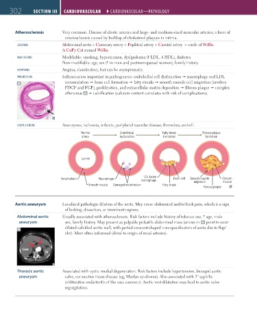
PROGRESSION Inflammation important in pathogenesis: endothelial cell dysfunction macrophage and LDL
A accumulation foam cell formation fatty streaks smooth muscle cell migration (involves
PDGF and FGF), proliferation, and extracellular matrix deposition fibrous plaque complex
atheromas A calcification (calcium content correlates with risk of complications).
COMPlICATIONS Aneurysms, ischemia, infarcts, peripheral vascular disease, thrombus, emboli.
Normal Endothelial Fatty streak Fibrous plaque
artery dysfunction formation formation
Lumen
LDL-laden
Endothelium Macrophage Foam cell Smooth muscle Smooth
macrophage migration muscle
Smooth muscle Damaged endothelium Fatty streak
Fibrous plaque
Aortic aneurysm Localized pathologic dilation of the aorta. May cause abdominal and/or back pain, which is a sign
of leaking, dissection, or imminent rupture.
Abdominal aortic Usually associated with atherosclerosis. Risk factors include history of tobacco use, age, male
aneurysm sex, family history. May present as palpable pulsatile abdominal mass (arrows in A point to outer
dilated calcified aortic wall, with partial crescent-shaped non-opacification of aorta due to flap/
A
clot). Most often infrarenal (distal to origin of renal arteries).
Liver
Sp
Thoracic aortic Associated with cystic medial degeneration. Risk factors include hypertension, bicuspid aortic
aneurysm valve, connective tissue disease (eg, Marfan syndrome). Also associated with 3° syphilis
(obliterative endarteritis of the vasa vasorum). Aortic root dilatation may lead to aortic valve
regurgitation.
FAS1_2019_07-Cardio.indd 302 11/7/19 4:24 PM