
Page 352 - First Aid for the USMLE Step 1 2020, Thirtieth edition [MedicalBooksVN.com]_Neat
P. 352
308 SECTION III CARDIOvASCuLAR ``CARdIOvASCulAR—PATHOlOGY CARDIOvASCuLAR ``CARdIOvASCulAR—PATHOlOGY
Cardiomyopathies
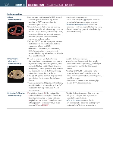
Dilated Most common cardiomyopathy (90% of cases). Leads to systolic dysfunction.
cardiomyopathy Often idiopathic or familial (eg, due to Dilated cardiomyopathy A displays eccentric
mutation of TTN gene encoding the hypertrophy (sarcomeres added in series).
A
sarcomeric protein titin). Takotsubo cardiomyopathy: broken heart
Other etiologies include drugs (eg, alcohol, syndrome—ventricular apical ballooning likely
cocaine, doxorubicin), infection (eg, coxsackie due to increased sympathetic stimulation (eg,
RV LV
B virus, Chagas disease), ischemia (eg, CAD), stressful situations).
systemic conditions (eg, hemochromatosis,
sarcoidosis, thyrotoxicosis, wet beriberi),
peripartum cardiomyopathy.
Findings: HF, S3, systolic regurgitant murmur,
dilated heart on echocardiogram, balloon
appearance of heart on CXR.
+
Treatment: Na restriction, ACE inhibitors,
β-blockers, diuretics, mineralocorticoid
receptor blockers (eg, spironolactone), digoxin,
ICD, heart transplant.
Hypertrophic 60–70% of cases are familial, autosomal Diastolic dysfunction ensues.
obstructive dominant (most commonly due to mutations Marked ventricular concentric hypertrophy
cardiomyopathy in genes encoding sarcomeric proteins, such (sarcomeres added in parallel) B , often septal
as myosin binding protein C and β-myosin predominance. Myofibrillar disarray and
B
heavy chain). Causes syncope during exercise fibrosis.
and may lead to sudden death (eg, in young Physiology of HOCM—asymmetric septal
athletes) due to ventricular arrhythmia. hypertrophy and systolic anterior motion of
RV Findings: S4, systolic murmur. May see mitral mitral valve outflow obstruction dyspnea,
LV
regurgitation due to impaired mitral valve possible syncope.
closure. Other causes of concentric LV hypertrophy:
Treatment: cessation of high-intensity athletics, chronic HTN, Friedreich ataxia.
2+
use of β-blocker or non-dihydropyridine Ca
channel blockers (eg, verapamil). ICD if
syncope occurs.
Restrictive/infiltrative Postradiation fibrosis, Löffler endocarditis, Diastolic dysfunction ensues. Can have low-
cardiomyopathy Endocardial fibroelastosis (thick fibroelastic voltage ECG despite thick myocardium
tissue in endocardium of young children), (especially in amyloidosis).
Amyloidosis, Sarcoidosis, Hemochromatosis Löffler endocarditis—associated with
(although dilated cardiomyopathy is more hypereosinophilic syndrome; histology shows
common) (Puppy LEASH). eosinophilic infiltrates in myocardium.
FAS1_2019_07-Cardio.indd 308 11/7/19 4:24 PM