Page 349 - First Aid for the USMLE Step 1 2020, Thirtieth edition [MedicalBooksVN.com]_Neat
P. 349

CARDIOvASCuLAR  ``CARdIOvASCulAR—PATHOlOGY      CARDIOvASCuLAR  ``CARdIOvASCulAR—PATHOlOGY            SECTION III      305


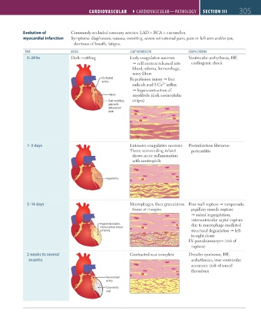
                  Evolution of           Commonly occluded coronary arteries: LAD > RCA > circumflex.
                  myocardial infarction  Symptoms: diaphoresis, nausea, vomiting, severe retrosternal pain, pain in left arm and/or jaw,
                                          shortness of breath, fatigue.

                   TIME                  GROSS                       lIGHT MICROSCOPE           COMPlICATIONS
                    0–24 hr              Dark mottling               Early coagulative necrosis   Ventricular arrhythmia, HF,
                                                                      Ž cell content released into   cardiogenic shock
                                                                      blood; edema, hemorrhage,
                                                                      wavy fibers
                                                       Occluded      Reperfusion injury Ž free
                                                       artery
                                                                                    2+
                                                                      radicals and  Ca  influx
                                                                      Ž hypercontraction of
                                                          Infarct     myofibrils (dark eosinophilic
                                                          Dark mottling;   stripes)
                                                          pale with
                                                          tetrazolium
                                                          stain


                    1–3 days                                         Extensive coagulative necrosis  Postinfarction fibrinous
                                                                     Tissue surrounding infarct   pericarditis
                                                                      shows acute inflammation
                                                                      with neutrophils


                                                         Hyperemia


                    3–14 days                                        Macrophages, then granulation  Free wall rupture Ž tamponade;
                                                                      tissue at margins          papillary muscle rupture
                                                                                                 Ž mitral regurgitation;
                                                                                                 interventricular septal rupture
                                                       Hyperemic border;                         due to macrophage-mediated
                                                       central yellow-brown
                                                       softening                                 structural degradation Ž left-
                                                                                                 to-right shunt
                                                                                                LV pseudoaneurysm (risk of
                                                                                                 rupture)
                    2 weeks to several                               Contracted scar complete   Dressler syndrome, HF,
                     months                                                                      arrhythmias, true ventricular
                                                                                                 aneurysm (risk of mural
                                                                                                 thrombus)
                                                         Recanalized
                                                         artery
                                                         Gray-white
                                                         scar


          FAS1_2019_07-Cardio.indd   305                                                                                11/7/19   4:24 PM
   344   345   346   347   348   349   350   351   352   353   354