Page 350 - First Aid for the USMLE Step 1 2020, Thirtieth edition [MedicalBooksVN.com]_Neat
P. 350

306        SECTION III    CARDIOvASCuLAR  ``CARdIOvASCulAR—PATHOlOGY                                                                                            CARDIOvASCuLAR  ``CARdIOvASCulAR—PATHOlOGY


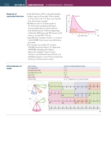
               Diagnosis of          In the first 6 hours, ECG is the gold standard.
               myocardial infarction  Cardiac troponin I rises after 4 hours (peaks   50  Troponin I
                                       at 24 hr) and is  for 7–10 days; more specific
                                       than other protein markers.                 10
                                     CK-MB rises after 6–12 hours (peaks at       Multiples of upper limit of normal
                                       16–24 hr) and is predominantly found
                                       in myocardium but can also be released      5
                                       from skeletal muscle. Useful in diagnosing   2    CK-MB                   Normal
                                       reinfarction following acute MI because levels   1
                                                                                                  4
                                                                                               3
                                                                                                      5
                                       return to normal after 48 hours.                 1  2  Days after MI onset  6  7  8
                                     Large MIs lead to greater elevations in troponin
                                       I and CK-MB. Exact curves vary with testing
                                       procedure.
                                     ECG changes can include ST elevation
                                       (STEMI, transmural infarct), ST depression
                                       (NSTEMI, subendocardial infarct),
                                       hyperacute (peaked) T waves, T-wave
                                       inversion, new left bundle branch block, and
                                       pathologic Q waves or poor R wave progression
                                       (evolving or old transmural infarct).


               ECG localization of    INFARCT LOCATION                        LEADS WITH ST-SEGMENT ELEVATIONS OR Q WAVES
               STEMI                  Anteroseptal (LAD)                      V –V 2
                                                                               1
                                      Anteroapical (distal LAD)               V –V 4
                                                                               3
                                      Anterolateral (LAD or LCX)              V 5 –V 6
                                      Lateral (LCX)                           I, aVL
                                      InFerior (RCA)                          II, III, aVF
                                      Posterior (PDA)                         V –V ,  ST depression in V –V  with tall R waves
                                                                                             1
                                                                                 9
                                                                               7
                                                                                               3
                                                         V6
                                                         V5   I             aVR           V1              V4
                                      Sternum
                                                     V4
                                                  V3
                                          V1   V2
                                                              II            aVL           V2              V5
                                       aVR             aVL

                                                        I     III           aVF           V3              V6


                                           III      II        II
                                               aVF


          FAS1_2019_07-Cardio.indd   306                                                                                11/7/19   4:24 PM
   345   346   347   348   349   350   351   352   353   354   355