
Page 351 - First Aid for the USMLE Step 1 2020, Thirtieth edition [MedicalBooksVN.com]_Neat
P. 351
CARDIOvASCuLAR ``CARdIOvASCulAR—PATHOlOGY CARDIOvASCuLAR ``CARdIOvASCulAR—PATHOlOGY SECTION III 307
Myocardial infarction complications
Cardiac arrhythmia Occurs within the first few days after MI. Important cause of death before reaching the hospital
and within the first 24 hours post-MI.
Postinfarction 1–3 days: friction rub.
fibrinous pericarditis
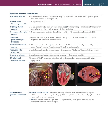
Papillary muscle 2–7 days: posteromedial papillary muscle rupture A risk due to single blood supply from posterior
rupture descending artery. Can result in severe mitral regurgitation.
Interventricular septal 3–5 days: macrophage-mediated degradation VSD O 2 saturation and pressure in RV.
rupture
Ventricular 3–14 days: free wall rupture contained by adherent pericardium or scar tissue B ; CO, risk of
pseudoaneurysm arrhythmia, embolus from mural thrombus.
formation
Ventricular free wall 5–14 days: free wall rupture C cardiac tamponade. LV hypertrophy and previous MI protect
rupture against free wall rupture. Acute form usually leads to sudden death.
True ventricular 2 weeks to several months: outward bulge with contraction (“dyskinesia”), associated with fibrosis.
aneurysm
Dressler syndrome Several weeks: autoimmune phenomenon resulting in fibrinous pericarditis.
LV failure and Can occur 2° to LV infarction, VSD, free wall rupture, papillary muscle rupture with mitral
pulmonary edema regurgitation.
A B C
Mitral valve
LA
RV LV
Pap
LV
Acute coronary Unstable angina/NSTEMI—Anticoagulation (eg, heparin), antiplatelet therapy (eg, aspirin)
syndrome treatments + ADP receptor inhibitors (eg, clopidogrel), β-blockers, ACE inhibitors, statins. Symptom control
with nitroglycerin and morphine.
STEMI—In addition to above, reperfusion therapy most important (percutaneous coronary
intervention preferred over fibrinolysis).
FAS1_2019_07-Cardio.indd 307 11/7/19 4:24 PM