
Page 432 - First Aid for the USMLE Step 1 2020, Thirtieth edition [MedicalBooksVN.com]_Neat
P. 432
388 seCtion iii Gastrointestinal ` gastrointestinal—PatHology Gastrointestinal ` gastrointestinal—PatHology
Lynch syndrome Previously called hereditary nonpolyposis colorectal cancer (HNPCC). Autosomal dominant
mutation of mismatch repair genes (eg, MLH1, MSH2) with subsequent microsatellite instability.
∼ 80% progress to CRC. Proximal colon is always involved. Associated with endometrial, ovarian,
and skin cancers.
Colorectal cancer
Diagnosis Iron deficiency anemia in males (especially > 50 years old) and postmenopausal females raises
A suspicion.
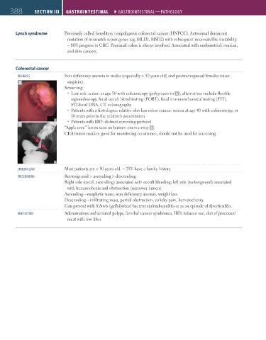
Screening:
Low risk: screen at age 50 with colonoscopy (polyp seen in A ); alternatives include flexible
sigmoidoscopy, fecal occult blood testing (FOBT), fecal immunochemical testing (FIT),
FIT-fecal DNA, CT colonography
Patients with a first-degree relative who has colon cancer: screen at age 40 with colonoscopy, or
10 years prior to the relative's presentation
Patients with IBD: distinct screening protocol
B
“Apple core” lesion seen on barium enema x-ray B .
CEA tumor marker: good for monitoring recurrence, should not be used for screening.
ePiDemiology Most patients are > 50 years old. ~ 25% have a family history.
Presentation Rectosigmoid > ascending > descending.
Right side (cecal, ascending) associated with occult bleeding; left side (rectosigmoid) associated
with hematochezia and obstruction (narrower lumen).
Ascending—exophytic mass, iron deficiency anemia, weight loss.
Descending—infiltrating mass, partial obstruction, colicky pain, hematochezia.
Can present with S bovis (gallolyticus) bacteremia/endocarditis or as an episode of diverticulitis.
risK FaCtors Adenomatous and serrated polyps, familial cancer syndromes, IBD, tobacco use, diet of processed
meat with low fiber.
FAS1_2019_09-Gastrointestinal.indd 388 11/7/19 4:42 PM