
Page 570 - First Aid for the USMLE Step 1 2020, Thirtieth edition [MedicalBooksVN.com]_Neat
P. 570
526 SecTioN iii Neurology aNd Special SeNSeS ` neurology—PAthology Neurology aNd Special SeNSeS ` neurology—PAthology
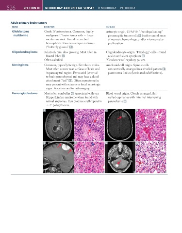
Adult primary brain tumors
tumor desCriPtion histology
Glioblastoma Grade IV astrocytoma. Common, highly Astrocyte origin, GFAP ⊕. “Pseudopalisading”
multiforme malignant 1° brain tumor with ~ 1-year pleomorphic tumor cells B border central areas
median survival. Found in cerebral of necrosis, hemorrhage, and/or microvascular
hemispheres. Can cross corpus callosum proliferation.
(“butterfly glioma” A ).
Oligodendroglioma Relatively rare, slow growing. Most often in Oligodendrocyte origin. “Fried egg” cells—round
frontal lobes C . nuclei with clear cytoplasm D.
Often calcified. “Chicken-wire” capillary pattern.
Meningioma Common, typically benign. Females > males. Arachnoid cell origin. Spindle cells
Most often occurs near surfaces of brain and concentrically arranged in a whorled pattern F ;
in parasagittal region. Extra-axial (external psammoma bodies (laminated calcifications).
to brain parenchyma) and may have a dural
attachment (“tail” E ). Often asymptomatic;
may present with seizures or focal neurologic
signs. Resection and/or radiosurgery.
Hemangioblastoma Most often cerebellar G. Associated with von Blood vessel origin. Closely arranged, thin-
Hippel-Lindau syndrome when found with walled capillaries with minimal intervening
retinal angiomas. Can produce erythropoietin parenchyma H.
2° polycythemia.
A B C D
E F G H
FAS1_2019_12-Neurol.indd 526 11/8/19 7:39 AM