
Page 583 - First Aid for the USMLE Step 1 2020, Thirtieth edition [MedicalBooksVN.com]_Neat
P. 583
Neurology aNd Special SeNSeS ` neurology—oPhthAlmology Neurology aNd Special SeNSeS ` neurology—oPhthAlmology SecTioN iii 539
Pupillary control
Miosis Constriction, parasympathetic:
1st neuron: Edinger-Westphal nucleus to ciliary ganglion via CN III
2nd neuron: short ciliary nerves to sphincter pupillae muscles
Short ciliary nerves shorten the pupil diameter.
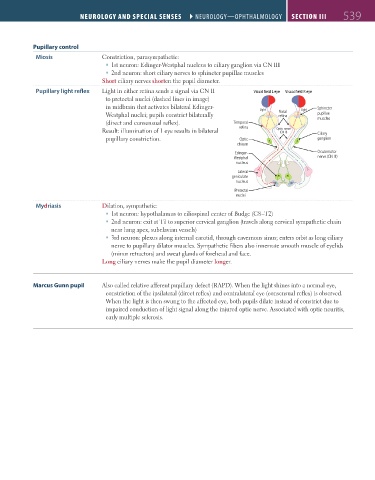
Pupillary light reflex Light in either retina sends a signal via CN II Visual field L eye Visual field R eye
to pretectal nuclei (dashed lines in image)
in midbrain that activates bilateral Edinger- Sphincter
Light Nasal Light
Westphal nuclei; pupils constrict bilaterally retina pupillae
(direct and consensual reflex). Temporal muscles
Result: illumination of 1 eye results in bilateral retina Optic nerve Ciliary
(CN II)
pupillary constriction. Optic ganglion
chiasm
Edinger- Oculomotor
Westphal nerve (CN III)
nucleus
Lateral
geniculate
nucleus
Pretectal
nuclei
Mydriasis Dilation, sympathetic:
1st neuron: hypothalamus to ciliospinal center of Budge (C8–T2)
2nd neuron: exit at T1 to superior cervical ganglion (travels along cervical sympathetic chain
near lung apex, subclavian vessels)
3rd neuron: plexus along internal carotid, through cavernous sinus; enters orbit as long ciliary
nerve to pupillary dilator muscles. Sympathetic fibers also innervate smooth muscle of eyelids
(minor retractors) and sweat glands of forehead and face.
Long ciliary nerves make the pupil diameter longer.
Marcus Gunn pupil Also called relative afferent pupillary defect (RAPD). When the light shines into a normal eye,
constriction of the ipsilateral (direct reflex) and contralateral eye (consensual reflex) is observed.
When the light is then swung to the affected eye, both pupils dilate instead of constrict due to
impaired conduction of light signal along the injured optic nerve. Associated with optic neuritis,
early multiple sclerosis.
FAS1_2019_12-Neurol.indd 539 11/8/19 7:39 AM