
Page 580 - First Aid for the USMLE Step 1 2020, Thirtieth edition [MedicalBooksVN.com]_Neat
P. 580
536 SecTioN iii Neurology aNd Special SeNSeS ` neurology—oPhthAlmology Neurology aNd Special SeNSeS ` neurology—oPhthAlmology
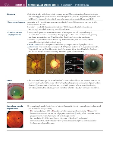
Glaucoma Optic disc atrophy with characteristic cupping (normal A versus thinning of outer rim of optic
nerve head B ), usually with elevated intraocular pressure (IOP) and progressive peripheral visual
field loss if untreated. Treatment is through pharmacologic or surgical lowering of IOP.
Open-angle glaucoma Associated with age, African-American race, family history. Painless, more common in US.
Primary—cause unclear.
Secondary—blocked trabecular meshwork from WBCs (eg, uveitis), RBCs (eg, vitreous
hemorrhage), retinal elements (eg, retinal detachment).
Closed- or narrow- Primary—enlargement or anterior movement of lens against central iris (pupil margin)
angle glaucoma obstruction of normal aqueous flow through pupil fluid builds up behind iris, pushing
peripheral iris against cornea C and impeding flow through trabecular meshwork.
Secondary—hypoxia from retinal disease (eg, diabetes mellitus, vein occlusion) induces
vasoproliferation in iris that contracts angle.
Chronic closure—often asymptomatic with damage to optic nerve and peripheral vision.
Acute closure—true ophthalmic emergency. IOP pushes iris forward angle closes abruptly.
Very painful, red eye D, sudden vision loss, halos around lights, frontal headache, fixed and
mid-dilated pupil, nausea and vomiting. Mydriatic agents contraindicated.
A B C D
Normal
Normal Cupping Angle closure Acute angle closure
Uveitis Inflammation of uvea; specific name based on location within affected eye. Anterior uveitis: iritis;
A posterior uveitis: choroiditis and/or retinitis. May have hypopyon (accumulation of pus in anterior
chamber A ) or conjunctival redness. Associated with systemic inflammatory disorders (eg,
sarcoidosis, rheumatoid arthritis, juvenile idiopathic arthritis, HLA-B27–associated conditions).
Age-related macular Degeneration of macula (central area of retina). Causes distortion (metamorphopsia) and eventual
degeneration loss of central vision (scotomas).
Dry (nonexudative, > 80%)—Deposition of yellowish extracellular material (“Drusen”) in
A
between Bruch membrane and retinal pigment epithelium A with gradual in vision. Prevent
progression with multivitamin and antioxidant supplements.
Wet (exudative, 10–15%)—rapid loss of vision due to bleeding 2° to choroidal
neovascularization. Treat with anti-VEGF (vascular endothelial growth factor) injections
(eg, bevacizumab, ranibizumab).
FAS1_2019_12-Neurol.indd 536 11/8/19 7:39 AM