
Page 581 - First Aid for the USMLE Step 1 2020, Thirtieth edition [MedicalBooksVN.com]_Neat
P. 581
Neurology aNd Special SeNSeS ` neurology—oPhthAlmology Neurology aNd Special SeNSeS ` neurology—oPhthAlmology SecTioN iii 537
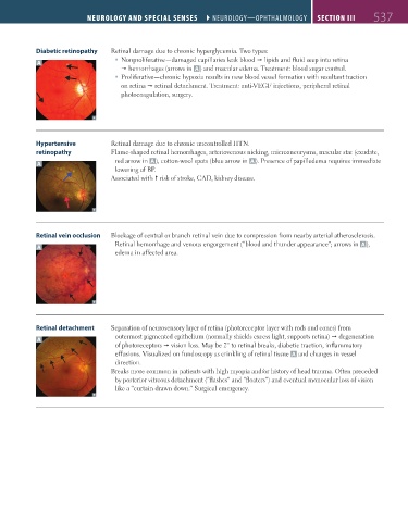
Diabetic retinopathy Retinal damage due to chronic hyperglycemia. Two types:
Nonproliferative—damaged capillaries leak blood lipids and fluid seep into retina
A
hemorrhages (arrows in A ) and macular edema. Treatment: blood sugar control.
Proliferative—chronic hypoxia results in new blood vessel formation with resultant traction
on retina retinal detachment. Treatment: anti-VEGF injections, peripheral retinal
photocoagulation, surgery.
Hypertensive Retinal damage due to chronic uncontrolled HTN.
retinopathy Flame-shaped retinal hemorrhages, arteriovenous nicking, microaneurysms, macular star (exudate,
red arrow in A ), cotton-wool spots (blue arrow in A ). Presence of papilledema requires immediate
A
lowering of BP.
Associated with risk of stroke, CAD, kidney disease.
Retinal vein occlusion Blockage of central or branch retinal vein due to compression from nearby arterial atherosclerosis.
Retinal hemorrhage and venous engorgement (“blood and thunder appearance”; arrows in A ),
A
edema in affected area.
Retinal detachment Separation of neurosensory layer of retina (photoreceptor layer with rods and cones) from
A outermost pigmented epithelium (normally shields excess light, supports retina) degeneration
of photoreceptors vision loss. May be 2° to retinal breaks, diabetic traction, inflammatory
effusions. Visualized on fundoscopy as crinkling of retinal tissue A and changes in vessel
direction.
Breaks more common in patients with high myopia and/or history of head trauma. Often preceded
by posterior vitreous detachment (“flashes” and “floaters”) and eventual monocular loss of vision
like a “curtain drawn down.” Surgical emergency.
FAS1_2019_12-Neurol.indd 537 11/8/19 7:39 AM